Download
1 / 23
230 likes | 398 Vues
HN5. PARA PHARYNGEAL GERM CELL TUMOR. R. AOUINI, S. KOUKI, A. HRICHI , I. GANZOUI, M. LANDOULSI , S. BOUGUERRA, Y. AROUS, H. BOUJEMAA, N. BEN ABDALLAH. INTRODUCTION:.

E N D
HN5 PARA PHARYNGEAL GERM CELL TUMOR R. AOUINI, S. KOUKI, A. HRICHI , I. GANZOUI, M. LANDOULSI , S. BOUGUERRA, Y. AROUS, H. BOUJEMAA, N. BEN ABDALLAH
INTRODUCTION: • Germ cell tumors (GCTs) are a heterogeneous group of lesions which arise in patients of all ages; they occur most frequently in the gonads and are relatively rare in other sites. • Both computed tomography and magnetic resonance imaging (MRI) are highly sensitive in the detection of these tumors. • In this case we report an uncommon localization of a germ cell tumor which is the Para pharyngeal space.
MATERIALS AND METHODS: • A 5 year old boy without any medical history complaining of facial paralysis which doesn’t respond to symptomatic treatments and persisting for more than 2 weeks. • A CT scan was performed with axial, frontal and sagittal reconstruction before and after injection of a contrast agent.
RESULTS: • Physical examination revealed only a peripheral facial paralysis. • Laboratory studies disclosed no abnormalities in the hemogram or urinanalysis. • Serumα-fetoproteinlevelwaselevated. • Human chorionic gonadotropin (HCG) determination was normal.
UnenhancedCT scan shows a welll-circumscribedhomogeneous and isodense mass measuring 35 x 20 mm of diametresin the right parapharyngealspacerepressing the medialpterygoidmuscle and filling the Eustachian tube and the fossa of Rosen muller. This lesionextends to the infra temporal space.
This mass shows a heterogeneous enhancement after contrast administration, the low-attenuation area corresponds to necrosis.The mass represses the neck vessels.
This mass alsoinvades the right temporal lobe with the presence of hypoattenuating unenhanced areas at CT corresponding to necrosis.
right middle ear is filled with integrity of the ossicularchain. No evidenceof vestibularor labyrinthic invasion. lysis of the petrous part of temporal bone responsible for a lesion of the facial nerve.
hyperdense images testify of the presence of bone destruction with small fragments detached
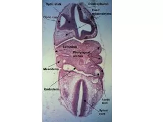
DISCUSSION: • Germ cell tumors are neoplasms arising from primordial germ cells and are composed of embryonic ectodermal, mesodermal, and endodermal tissue and/or extra-embryonic tissues, such as trophoblast and yolk sac. • Embryologicand histopathologic considerations suggest two different origins of extragonadal GCTs: metastases from gonadal GCTs and primary GCTs originating from migrated primordial germ cells. • Among the various histologic subtypes of GCTs benign teratomas are most frequently found[8].Overall, the prevalenceof nonseminomatous GCTs is much less thanthat of seminomas.
GCTs mainly occur in the gonads and localization at extragonadalsites(e i :no evidence of a primary tumor in either the testes or the ovaries.[3] ) is uncommon. • Most extragonadal GCTs occur in the median line of the human body: the anterior mediastinum, sacrococcygealregion, pineal gland, andneurohypophysis are common sites. • In extracranial head and neck regions, they are extremely rare: 5%[4,5,6,7] • the histologicfeatures of each tumor variety are similar whereveritoccurs.[9]
Site distribution and frequency of germ cell tumors (children < 15 years).[1]
The age at diagnosis shows a bimodal peak with an increased incidence in the first four years of life and then from second to fourth decade of life.[2] • Abnormal karyotypes, and Conditions such as male cryptorchidism, aniridia-Wilms’ association, sacral agenesis and males with Russell-Silver syndrome have an increased risk of GCTs.[2]
In general, GCTs tend to occur as indolent masses, and clinical symptoms are mostly related to local dysfunction by tumor growth, the paralysisof the cranial nerves can be the first manifestation especially the V, VII, IX, X, XII. • In our case peripheral facial paralysiswas the onlyclinical manifestation leading to perform a facial and cerebral CT scan. • Secretion of AFP and less commonly ß-HCG can be important in diagnosis, assessing treatment response and post-treatment surveillance (CSF measurement in suspected intracranial GCTs is mandatory.)
Radiologicfindings varies depending on the histologic type of tumor(seminoma, non seminomatoisGCTs, teratoma): • Seminomasproduce a bulky lobulated well marginated solid homogeneous with fibrovascularsepta(low-signal intensity bands on T2), mildly enhancing masses on CT[9,10], calcification are rarelyseen. MRI appearanceis non specific. Local invasion isuncommon, howeverlymphnode metatases are oftenpresentatpresentation[9].
Teratomas may be mature (benign) or immature (with variable malignant potential)[9]: • Malignant teratomas usually have a large solid component and a mixed histology, with the presence of elements of other germ cell tumors. • Mature teratomas appear as well-defined lobulated heterogeneous solid or cystic masses. Calcifications are found in ¼ to 1/3 of cases.
Non seminomatous GCTs are usually large, lobulated, heterogeneous and infiltrating masses with irregular margins containing calcification, hemorrhage, and necrosis. MRI allowbettercharacterization of the different components of the tumorsuch as areas of hemmoragewhichappear as hyperintense on T1 weighted images, cysts and necrotic areas as hyperintense on T2-weighted images. Invasion of adjacent organsiscommon[9,10].
The diagnosis is based on biopsy and assay of specific serum markers including alpha-foeto protein (A.F.P). In our case histology shows a tumor proliferation associated with the classic Schiller-Duval bodies which are characterizedby a central blood vessel covered by tumor cells and separated from an outer rim of tumor cells by a clear space. • Histopathologically Schiller Duval bodies when present are pathognomonic of the Yolk sac tumor.
The treatement and the prognosis for GCTs depends on the histologic subtype: • Treatment of mature teratomas is completed only by surgical removal. • Seminomas, are very sensitive to radiation and chemotherapy. • Non seminomatous GCTs should benefit of surgical removal when its possible, in association with a multi-agent chemotherapy.[8] Theirprognosisispoor due to theiraggressivity and deep location responsiblefor delayeddiagnosis.
CONCLUSION: • Extragonadal germ cell tumors have a variable clinical course, with the potential for aggressive behavior and widespread metastases. • The imaging characteristics of these tumors are nonspecific, and in combination with other clinical data, including tumor markers, should always lead to consideration of extragonadalgerm cell tumors and the definitivediagnosis of extragonadalGCTs requires a biopsy.
BIBLIOGRAPHIE: • 1-Nadira Mamoon,1 Sadaf Ali Jaffri,2 Fazal Ilahi,3 Kamil Muzaffar,4 Yasir Iqbal,5 Noreen Akhter,6 Humaira Nasir,7 Imran Nazir Ahmad8 Yolk sac tumour arising in mature teratoma in the parapharyngeal spaceJ Pak Med Assoc. • 2-Matthew Jonathan Murray, James Christopher Nicholson, Germ cell tumors in children and adolescents,Paediatrics and child health 20:3. • 3-Gabriele Calaminus, Catherine Patte Germ Cell Tumors in Children and Adolescents international society of pediatriconcology. • 4-Kusumakumari P., Geetha N., Chellam VG., Nair MK. Endodermal sinus tumorsin the headand neck region. Med PediatrOncol. 1997; 29(4):303-7. • 5Lack EE. Extragonadal germ cell tumors of the head and neck region: review of 16 cases. Hum Pathol. 1985 ; 16(1):56-64. • 6-Dehner LP., Mills A., Talerman A., Billman GF., Krous HF., Platz CE. Germ cell neoplasms of head and neck soft tissues: a pathologic spectrum of teratomatous and endodermal sinus tumors. Hum Pathol. 1990; 21(3):309-18.
7-I. Gassab, A. Hamroun, K. Harrathi, A. Hizem, F. Ben Mahmoud,F. El Kadhi, A. Moussa*, CH. Hafsa**, J. Koubaa, A.GassabTumeur germinale de l’espace parapharyngé: a propos d’un cas. J. TUN ORL - N° 18 JUIN 2007 • 8-Gerrit-Jan Westerveld, MD, Jasper J. Quak, MD, PhD, DorineBresters, MD, PhD, Christian M. Zwaan, MD,Paul Van Der Valk, MD, PhD, and Charles R. Leemans, MD, PhD, Endodermal sinus tumor of the maxillary sinus. Otolaryngology–Head and Neck Surgery June 2001. • 9-Atul B. Shinagare, JyothiP. Jagannathan, Nikhil H. Ramaiya, Matthew N. Hall, Annick D. Van den AbbeeleAdultExtragonadalGermCellTumors. AJR:195, October2010. • 10-Teruko Ueno, MD, YumikoOishi Tanaka, MD, MichioNagata, MD HajimeTsunoda, MD, IzumiAnno, MD, Shigemi Ishikawa, MD Koji Kawai, MD, YujiItai, MD, Spectrum of GermCellTumors: FromHead to Toe, RadioGraphics2004.