Download
1 / 32
320 likes | 479 Vues
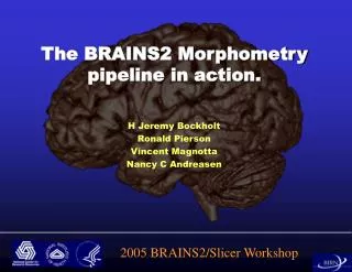
The BRAINS2 Morphometry pipeline in action. H Jeremy Bockholt Ronald Pierson Vincent Magnotta Nancy C Andreasen. 2005 BRAINS2/Slicer Workshop. Depends on the question that you are asking - Volumetric Analysis: How big is it? What kind of tissue is there and how much of it?

E N D
The BRAINS2 Morphometry pipeline in action. H Jeremy Bockholt Ronald Pierson Vincent Magnotta Nancy C Andreasen 2005 BRAINS2/Slicer Workshop
Depends on the question that you are asking - Volumetric Analysis: How big is it? What kind of tissue is there and how much of it? Morphometric Analysis: What is the size and shape of the brain or of its structures? Other types – DTI and Spectroscopy: What other static characteristics can we measure? White matter direction and coherence, and concentrations of biologically significant chemicals Use of ROIs for functional image analysis Reasons Structural Analysis
Volumetric Analysis: Measure volumes of tissue in gross regions of the brain Automate the process to make it possible to handle large volumes of scans Remove or minimize effects of different raters and rater fatigue or drift Create a set of images that will be useful for future work – measurement of other structures via manual tracing, etc. Basic Goals of Standard Workup:
Acquire MR Images Resample/Coregister MR Images Tissue Classification Neural Network Structure Identification Measure Volumes Surface Generation Surface Measurements Standard Workup Overview
Each site acquires T1 and either T2 or PD. Iowa acquires single NEX=2 T1 and Nex=3 T2. Other 3 sites acquire multiple NEX=1 scans. QA review at each site before uploading to SRB and after downloading at Iowa prior to processing. Image Acquisition
T1 images are realigned in a standard orientation. The standard orientation calls for lining up the interhemispheric fissure. This sets the alignment in the coronal and axial planes. In addition, the anterior commisure and posterior commisure are used for the horizontal orientation in the sagittal plane. Manual Resampling
All other images are coregistered to the manually reoriented T1 by use of AIR or Mutual Information coregistration. For those sites acquiring multiple NEX=1 scans, after coregistration all of the scans in each modality are averaged together to produce an image with better CNR. When fitting is complete each image is resampled to a new orientation and a resolution of 0.5 mm cubic voxels. Point to point correspondence with any given set of coordinates referring to the same point in all of the images Coregistration
Define a Talairach-based atlas for the each scan individually Landmarks used Right-most extent of the brain Left-most extent of the brain Anterior-most extent of the brain Posterior-most extent of the brain Superior-most extent of the brain Inferior-most extent of the temporal lobe AC and PC locations Talairach Bounds
Talairach Atlas Talairach atlas coordinate system Resampled image with overlaid Talairach coordinate system
Talairach Atlas warped onto current brain. Various "boxes" assigned to various regions Measure volumes of labelled brain regions Talairach Regions Talairach Boxes Cyan - Frontal Blue - Temporal Green -Parietal Red - Occipital Pink - Cerebellum Yellow - Subcortical Gray - Brainstem
Tissue characteristics in a scan are determined by sampling for three possible classifications – gray matter, white matter and CSF. Blood is traced. Using these “training classes”, create a set of rules to classify each voxel in the image. Multiple modalities used, makes it possible to define the edge of the surface CSF. How do we know what type of tissue each voxel is?
Randomly choose 2x2x2 mm plugs Keep “pure” plugs - those with sufficiently low variance K-means cluster the plugs to assign them to GM, WM, or CSF Generate discriminant functions based on tissue assigned plugs Apply discriminant functions to the entire image Tissue Classification
The basis for all subsequent steps in standard workup Neural network structure identification Cortical surface generation Image normalization and enhancement Defines the tissue type at each voxel in the image Continuous classification - Multiple tissue types per voxel Discrete classification - Single tissue type per voxel Tissue Classification
Tissue classified image is coded on an 8 bit scale Other = 0, Blood = 1 Pure CSF = 10, Pure GM = 130, Pure WM = 250 Partial volume between CSF-GM and GM-WM Discrete image generated from continuous image using the following formula. CSF:10£x£70, GM: 70 < x £190 WM: 190<x£ 250 Classified Images
Artificial Neural Network used to define "Brain" Trained from manual traces Uses a standard, 3 layer, fully connected neural network Trained using back-propagation Inputs Signal intensity within a spherical region of the voxel Probability information Spatial location information Definition of the "BRAIN"
Most regional cutouts are reliable before editing Output of neural network trimmed for validity ROI Editing
Generate measures both for continuous and discrete images In general, discrete data has been used Regional measures partitioned into GM, WM, CSF, blood and other. Measurements made for total and internal CSF Can compute surface CSF based on these results Measurements corrected for signal inhomogeniety Tissue-Classified Volumes
In each region the volumes are measured for GM, WM, CSF, blood and other (unclassified) Frontal, temporal, parietal and occipital lobes Subcortical, cerebellum and brainstem Ventricles Add and subtract variables to create measures of interest Tissue-Classified Volumes
Use these ROIs to define masks which represent exclusion regions for surface generation – “the surface can’t go here.” Use a marching cubes type algorithm (Wyvill) to define the 130 isosurface in the image. Parametric center of GM Helps avoids the buried cortex problem Limit search space to side of interest Start out on the correct side of the hemisphere traces Keep the largest connected surface Repeat for other side Surface Generation Algorithm
Curvature Look at current triangle wrt local neighborhood of triangles up to 3 triangles away Determine if the current triangle is concave or convex Cortical depth Follow normal from center of triangle as well as each vertex Find shortest distance to 190 value (WM border) Algorithm Additions
Many, Many, Many variables ............ Measurements of Interest Surface Area (mm2): Gyral, Fundal, Total Curvature: Gyral or Fundal Thickness (mm): Gyral, Fundal, Total Measures obtained by Talairach boxes as well BE CAREFUL USING REGIONAL MEASURES Surface Measurements
Acquire MR Images Resample/Coregister MR Images Tissue Classification Definition of Brain Regional Structure Identification Volumetric Measurements Surface Generation Standard Workup Complete
Currently defines the following regions Caudate Putamen Thalamus Cerebellum Cerebellar lobes (warping) Hippocampus (requires editing) Globus Pallidus (requires editing) In the near future will use a warped method for all structures – more valid, less editing Will also add nucleus accumbens and amygdala Neural Network
Cerebellum Lobes Cerebellar Lobe Volumes: Uses landmark-based warp for semiautomated measurement of Lobes I through V (anterior lobe), Lobe VI and Crus I of VIIA (superior posterior lobe), Crus II of VIIA through Lobe X (inferior posterior lobe), and the central white matter and output nuclei(corpus medullare).
Manual Tracing Tools provided in BRAINS2 facilitate accurate tracing using multiple images and views. Useful for accurate placement of ROIs for DTI, functional image analysis. Can create spheres, cubes around a point Convert to code image – warp, coregister, import into SPM, etc. Parcellation of cortical surface
Create rigorously valid cortical lobe definitions by warping a template brain to individual’s scan. Other high-dimensional, non-linear warp projects to analyze shape FreeSurfer – semiautomated cortical parcellation Future Methods Available
Talairach Atlas – the space which the brain occupies is broken up into boxes, and each box is labeled with what region it belongs to. Create an atlas for each scan (based on the Talairach atlas) that does a good job of defining brain regions Also need a way to define what is brain and what is not Automated Regional Measures
What about the cerebellum? Not included in Talairach Atlas We have added two additional boxes to the inferior aspect of the Talairach atlas to include the cerebellum Used for automated gross regional measures Provides a coordinate system for structure probability Talairach Atlas II