Download
1 / 19
200 likes | 381 Vues
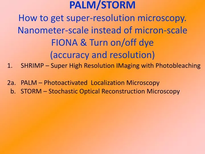
PALM/STORM How to get super-resolution microscopy. Nanometer-scale instead of micron-scale FIONA & Turn on/off dye (accuracy and resolution). SHRIMP – Super High Resolution IMaging with Photobleaching 2a. PALM – Photoactivated Localization Microscopy

E N D
PALM/STORM How to get super-resolution microscopy. Nanometer-scale instead of micron-scale FIONA & Turn on/off dye (accuracy and resolution) • SHRIMP – Super High Resolution IMaging with Photobleaching • 2a. PALM – Photoactivated Localization Microscopy • b. STORM – Stochastic Optical Reconstruction Microscopy
How fine can you see? The Limits of Microscopy For visible microscopy, Resolution is limited to ~250 nm Ernst Abbe & Lord Rayleigh Ernst Abbe Recent microscopy: 1-100 nm, Here we present techniques which are able to get super-accuracy (1.5 nm) and/or super-resolution (<10 nm, 35 nm)
center width FIONA: Diffraction limited spot: Single Molecule Sensitivity Accuracy of Center = width/ S-N = 250 nm / √104= ~2.5 nm = ± 1.25nm Width of l/2 ≈250 nm Enough photons (signal to noise)…Center determined to ~1.3 nm Dye lasts 5-10x longer -- typically ~30 sec- 1 min. (up to 4 min) Start of high-accuracy single molecule microscopy Thompson, BJ, 2002; Yildiz, Science, 2003
Super-Resolution: Nanometer Distances between two (or more) dyes SHRImP Super High Resolution IMaging with Photobleaching In vitro
132.9 ± 0.93 nm 72.1 ± 3.5 nm 8.7 ± 1.4 nm Super-Resolution: Nanometer Distances between two (or more) dyes SHRImP Super High Resolution IMaging with Photobleaching Distance can be found much more accurately than width (250 nm) Resolution now: Between 2-5 molecules: <10 nm (Gordon et al.; Qu et al, PNAS, 2004) Next slides gSHRIMP: > 5-15 molecules ~ 20-100 nm Via 2-photon: ~ 35 nm (next time) In vitro
Regular Microtubules (In vitro) Image • Take regular Image. • Then one fluorophore photobleaches. • Subtract off, get high resolution, repeat. Imaging resolution 300 nm 1000 nm Rhodamine-labeled microtubules, TIR Actual 24 nm; Measured 300nm 60
Regular- resolution image Super- Microtubules in a COS-7 cell Spots localized versus frame 2 um • 500 nm Standard FWHM: 560 ± 20 nm • 500 nm gSHRImP FWHM: 96.5 ± 1 nm
STochastic Optical Reconstruction Microscopy PhotoActivation Localization Microscopy STORM & PALMMost Super-Resolution MicroscopyInherently a single-molecule technique Zhuang, 2007 Science Betzig, 2006 Science Huang, Annu. Rev. Biochem, 2009 Cy3-Alexa 647 2-color secondary antibodies Cy2-Alexa 647
Make dyes blink with dye-pairs Photo-switching of Cy3-Cy5, 5.5, 7 532 nm 657 nm always on– excites Cy-dye & turns it off Cy3-Cy5 on DNA, antibody
Fig. 2. Three-dimensional STORM imaging of microtubules in a cell. Conventional indirect immunofluorescence image of microtubules 3D section (color coded) C-E zoom in of box in B B Huang et al. Science 2008;319:810-813 Published by AAAS
3D Movie B Huang et al. Science 2008;319:810-813
Fig. 3. Three-dimensional STORM imaging of clathrin-coated pits in a cell. Magnified View 2D STORM of same region (all z’s) Conventional direct immunofluorescence image of clathrin x-y cross section Magnified View B Huang et al. Science 2008;319:810-813 Published by AAAS
PALM Use photoswitchable GFP Numerous sparse subsets of photoactivatable fluorescent protein molecules were activated, localized (to ~2 to 25 nanometers), and then bleached. The aggregate position information from all subsets was then assembled into a super-resolution image. Fig. 1. The principle behind PALM. A sparse subset of PA-FP molecules that are attached to proteins of interest and then fixed within a cell are activated (A and B) with a brief laser pulse at λact = 405 mm and then imaged at λexc = 561 mm until most are bleached (C). This process is repeated many times (C and D) until the population of inactivated, unbleached molecules is depleted. Summing the molecular images across all frames results in a diffraction-limited image (E and F). E Betzig et al. Science 2006;313:1642-1645 Published by AAAS
1 mm 1 mm 1 mm Correlative PALM-EM imaging TIRF PALM EM Mitochondrial targeting sequence tagged with mEOS Patterson et al., Science 2002
Photo-active GFP G. H. Patterson et al., Science 297, 1873 -1877 (2002) We report a photoactivatable variant of GFP that, after intense irradiation with 413-nanometer light, increases fluorescence 100 times when excited by 488-nanometer light and remains stable for days under aerobic conditions Native= filled circle Photoactivated= Open squares T203H GFP: PA-GFP Wild-type GFP
Photoactivation and imaging in vitro. G. H. Patterson et al., Science 297, 1873 -1877 (2002)
“Regular” dyes blink (Cy5,…)simultaneously excited with 514, 647