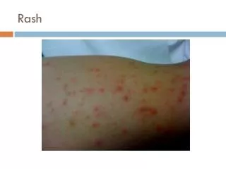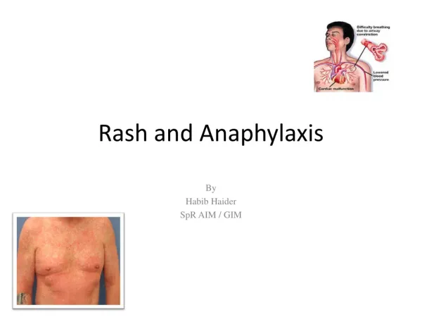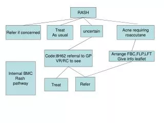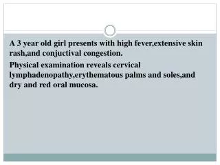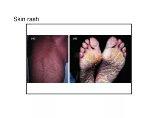
Fever and Rash
Fever and Rash. DR.Rezai MS Pediatrics infectious disease sub specialist. ماکول. ( اکیموز ). بثورات ماکولوپاپولر. پاپول . بثورات ماکولوپاپولر با توزیع محیطی . بثورات ماکولوپاپولر با توزیع مرکزی . وزیکول. پوستول . بول (طاول). ندول . (اریتم ندولار). اولسر نکروتیک .


Fever and Rash
E N D
Presentation Transcript
Fever and Rash DR.Rezai MS Pediatrics infectious disease sub specialist
ماکول ( اکیموز )
بثورات ماکولوپاپولر با توزیع محیطی بثورات ماکولوپاپولر با توزیع مرکزی
ندول (اریتم ندولار)
Classic Childhood Exanthems • Measles • Scarlet fever • Rubella (“German measles”) • Atypical scarlet fever • Erythema Infectiosum • Roseola
Adenovirus Anthrax Mononucleosis Colorado tick fever Mumps Cat-scratch fever Rat-bite fever Rocky Mountain spotted fever Relapsing fever Meningococcemia Typhus Hand-foot-mouth disease Today, dozens of exanthems are recognized:
Measles • Measles virus is a paramyxovirus • Paramyxoviruses : • Enveloped virus, ssRNA genome as a single piece. • The family includes parainfluenza virus, mumps virus, measles virus and respiratory syncytial virus. • Parainfluenza and mumps virus have a surface hemagglutinin and neuraminidase, while measles have a hemagglutinin, but not neuraminidase. • The virion structure includes: • Spikes • F protein • Matrix protein M, below the envelope • Only one serotype
#1- Measles • Virus: Rubeola • Demographics Winter or spring Infancy to young adulthood 8- to 12-day incubation Epidemics until 96% immunized • Prodrome 2–4 days. High fever, cough, coryza, conjunctivitis, photophobia, Koplik spots, lethargy, sneezing.
Measles Epidemiology • Reservoir Human • Transmission Respiratory Airborne • Temporal pattern Peak in late winter–spring • Communicability 4 days before to 4 days after rash onset
#1 MeaslesRash and Disease Enanthem: Koplik spots = gray pinheads, ring of erythema, buccal mucosa. 0.5–2d. Exanthem: erythematous blanching macules. Starts forehead, spreads downward Confluent by 72 hr Spares palms, and soles, 4–6 days. Toxic appearance.
#1- Measles • Diagnosis Leukopenia, IgG and IgM serologies, acute and convalescent titers • Treatment Symptomatic. Antipyretics. • In severe disease, vitamin A.
#1- Measles • Complications Otitis media, diarrhea, pneumonia (common in atypical rubeola). • Rarely, laryngo-tracheobronchitis, myocarditis, encephalitis. Subacute sclerosing panencephalitis
Complications • Giant cell pneumonia, more common in adults • Post-measles encephalitis • Subacute sclerosing panencephalitis (SSPE): • progressive and fatal degenerative disease • within the infected cells, there is a defective form of the virus which because it can not produce functional M protein, is not released as complete virus from the cells.
#1- MeaslesPrevention • Vaccinate all at 12–18 mo. • Two doses for 13 years and older. • Post-exposure vaccine if immuno-compromised • VZIG if pregnant, premature, or immunocompromised
Measles Vaccine • Composition Live virus • Efficacy 95% (range, 90%-98%) • Duration ofImmunity Lifelong • Schedule 2 doses
MMR VaccineContraindications and Precautions • Severe allergic reaction to vaccine component or following prior dose • Pregnancy • Immunosuppression • Moderate or severe acute illness • Recent blood product
#2- Scarlet Fever • Streptococcal, erythrogenic toxin. • Demographics 1 to 10 yr • Prodrome 2 to 4 days
Pathophysiology: • Streptococci are gram-positive cocci that grow in chains. They are classified by their ability to produce a zone of hemolysis on blood agar and by differences in carbohydrate cell wall components. • Streptococci may be alpha-hemolytic (partial hemolysis), beta-hemolytic (complete hemolysis), or gamma-hemolytic (no hemolysis). Most streptococci excrete hemolyzing enzymes and toxins. Erythrogenic toxins cause the rash of scarlet fever.
Background: • Scarlet fever (scarlatina) is an exotoxin-mediated disease arising from group A beta-hemolytic streptococcal infection. Ordinarily, scarlet fever evolves from a tonsillar pharyngeal focus, • Exotoxin-mediated streptococcal infections range from localized skin disorders (eg, bullous impetigo) to the systemic rash of scarlet fever to the uncommon but highly lethal streptococcal toxic shock syndrome.
#2- Scarlet Fever Rash and Disease • Strawberry tongue • Exudative pharyngitis • Generalized; spares palms and soles • Pinpoint papules • Desquamation of the tips of the fingers and toes
Sex: • Males and females are affected equally.
Age: • Peak incidence of scarlet fever occurs in persons aged 4-8 years. • By the time children are 10-years-old, 80% have developed lifelong protective antibodies against streptococcal pyrogenic exotoxins. • Scarlet fever is rare in children younger than 2 years, because of the presence of maternal antiexotoxin antibodies and lack of prior sensitization.
History: • The incubation period of streptococcal pharyngitis is usually 2-4 days. • Prodrome • Sore throat • Headache • Vomiting • Abdominal pain • Fever • The rash appears 12- 48 hours after onset of illness, first on the trunk and then extending rapidly over the entire body to finally involve the extremities. • Fever abates within 12-24 hours after initiation of antibiotic therapy.
Physical: • The patient usually appears moderately ill. • Fever • Tachycardia • Tonsils - Edematous, erythematous, and covered with a yellow, grey, or white exudate • Petechiae on the soft palate • Tender anterior cervical lymphadenopathy • Face - Flushed with perioral pallor
The exanthem is diffusely erythematous; but, in some patients, it is more palpable than visible. • Exanthem usually has the texture of coarse sandpaper, and the erythema blanches with pressure. • The skin can be pruritic but usually is not painful.
A few days following generalization of the rash, it becomes more intense along skin folds and produces lines of confluent petechiae known as the Pastia sign. These lines are caused by increased capillary fragility. • The rash begins to fade 3-4 days after onset, and the desquamation phase begins. This phase begins with flakes peeling from the face. Peeling from the palms and around the fingers occurs about a week later and lasts for about a month after onset of the disease.
The appearance of the tongue also has a characteristic course in scarlet fever. • During the first 2 days of the disease, the tongue has a white coat through which the red and edematous papillae project. This is referred to as a white strawberry tongue. • After 2 days, the tongue also desquamates, resulting in a red tongue with prominent papillae called the red strawberry tongue.
Lab Studies: • Throat culture remains the criterion standard for confirmation of group A streptococcal upper respiratory infection. • American Heart Association guidelines for prevention and treatment of rheumatic fever state that group A streptococci virtually always is found on throat culture during acute infection. • Throat cultures are approximately 90% sensitive for presence of group A beta-hemolytic streptococci in the pharynx. However, because a 10-15% carriage rate exists among healthy individuals, the presence of group A beta-hemolytic streptococci is not proof of disease. • To maximize sensitivity, proper obtaining of specimens is crucial. • Vigorously swab the posterior pharynx, tonsils, and any exudate with a cotton or Dacron swab under strong illumination, avoiding the lips, tongue, and buccal mucosa.
Direct antigen detection kits (ie, rapid antigen tests [RATs], strep screens) have been proposed to allow immediate diagnosis and prompt administration of antibiotics. • Kits are latex agglutination or a costlier enzyme-linked immunosorbent assay (ELISA). • Several trials of RAT kits report results of 78-100% specificity and 44-100% sensitivity compared to throat cultures. These studies usually were performed under laboratory conditions.
Streptococcal antibody tests are used to confirm previous group A streptococcal infection. • The most commonly available streptococcal antibody test is the antistreptolysin O test (ASLO test: antibodies to streptococcal extracellular products) . • Currently, streptococcal antibody tests during acute illness are not indicated. • These tests can provide confirmatory evidence of recent infection but have no value in acute infection. • They may be of value in patients suspected of having acute renal failure or acute glomerulonephritis.
Complete blood count • White blood cell (WBC) count in scarlet fever may increase to 12,000-16,000 per mm3, with a differential of up to 95% polymorphonuclear lymphocytes. • During the second week, eosinophilia, as high as 20%, can develop.