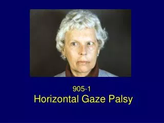
905-1
905-1. Horizontal Gaze Palsy. Left esotropia; fascicular sixth nerve palsy, left horizontal gaze palsy. Full horizontal gaze to the right with gaze evoked nystagmus. Vergence movements induced the right eye to cross the midline. Full Downgaze.

905-1
E N D
Presentation Transcript
905-1 Horizontal Gaze Palsy
Left esotropia; fascicular sixth nerve palsy, left horizontal gaze palsy
Full horizontal gaze to the right with gaze evoked nystagmus
Vergence movements induced the right eye to cross the midline
Impaired eye closure due to left facial palsy (Bell’s palsy)
Figure 1 Axial NECT scan shows a focal hemorrhage in the posterior pons and fourth ventricle. Patient with known breast cancer.
Figure 2 Sagittal NECT scan showing the rostral-caudal extent of the pontine hemorrhage
Ocular Motility Unilateral horizontal gaze palsy to the left that impaired saccades and pursuit Esotropia of the left eye Fascicular sixth nerve palsy Horizontal gaze full to the right, gaze evoked nystagmus
Ocular Motility Normal convergence, right eye induced to cross the midline Horizontal oculocephalic reflex, absent (Doll’s head maneuver) Vertical eye movements normal
Signs in Leigh and Zee’s Case The patient was unable to move her eyes to the right past the midline using either saccadic or pursuit eye movements Head rotation to the left, however, drove the eyes past the midline, but the right eye abducted incompletely Vergence movements induced the left eye to cross the midline Vertical eye movements were normal Gaze evoked nystagmus was present on looking to the left, with slow phases toward the midline The patient developed a fascicular sixth nerve palsy
Horizontal Gaze Palsy There are four theoretical possibilities to account for the ipsilateral horizontal gaze palsy due to a single unilateral lesion affecting 1. The ipsilateral paramedial pontine reticular formation (PPRF) only 2. The ipsilateral abducens nucleus (AN) alone 3. Both the ipsilateral PPRF and the AN, or when two lesions are involved 4. The motoneuron root fibers of the ipsilateral AN to the lateral rectus and the contralateral medial longitudinal fasciculus (MLF)
Figure 3 Horizontal section of the lower pons. 1) Basis pontis syndrome. 2) Internuclear ophthalmoplegia 3) Abducens nucleus syndrome 4) Caudal PPRF syndrome 5) One-and-a-half syndrome 6) Paramedian midbrain syndrome
Clinical Findings with PPRF Lesion Loss of horizontal saccades towards the side of the lesion Contralateral gaze deviation, in acute phase Gaze-evoked nystagmus on looking contralateral to the lesion
Clinical Findings with PPRF Lesion Impaired smooth pursuit and vestibular eye movements may be preserved or impaired Bilateral lesions cause total horizontal gaze palsy and slowing of vertical saccades
Clinical findings with lesion of the abducens nuclei Loss of all conjugate movements towards the side of the lesion – ipsilateral, horizontal gaze palsy Contralateral gaze deviation, in acute phase Vergence and vertical movements are spared
Clinical findings with lesion of the abducens nuclei In the intact hemifield of gaze, horizontal movements may be preserved, but ipsilaterally directed saccades are slow Horizontal gaze-evoked nystagmus on looking contralaterally Ipsilateral lower motoneuron facial palsy
Clinical distinction PPRF: AN at the bedside PPRF lesions rostral to abducens paralysis of saccades and pursuit, but the eyes can be driven to the side of the ipsilateral gaze palsy with vestibular stimulation by the oculocephalic reflex and/or cold calorics
Clinical distinction PPRF: AN at the bedside PPRF lesions at the level of abducens are associated with ipsilateral gaze palsy and loss of reflex vestibular (and tonic neck) movements This presumes that there is a critical synapse within the caudal PPRF for the vestibulo-ocular pathways or that the functional integrity of the PPRF at that level is necessary for vestibulo-ocular eye movements
Figure 5 Ocular motor control system. Combination of previous illustrations indicates saccadic (s), pursuit (P), and vestibular (VIII), inputs to PPRF and its output to the oculomotor nucleus (III) and the abducens nucleus (VI).
The PRF contains three types of saccade – related neurons Burst neurons (BN) Excitatory BN (EBN) create saccadic eye velocity commands (the pulse) Inhibitory BN (IBN) permit reciprocal innervation to occur Tonic neurons (TN) Part of the neural integrator that integrates eye velocity commands and holds position for gaze Pause neurons (PN) Exert a normal inhibitory influence upon saccadic burst neurons during periods of fixation
Figure 8 The motor circuit for horizontal saccades in the brainstem