AV Heart Blocks
AV Heart Blocks. Pauline Seydak, Cardiac Physiology Trainer N.I. AV Nodal Blocks (heart blocks). Disturbances of the conduction through the heart, occurring at the AV Node AV Node – damaged/diseased – delay or total block of impulses at the AV Node

AV Heart Blocks
E N D
Presentation Transcript
AV Heart Blocks Pauline Seydak, Cardiac Physiology Trainer N.I.
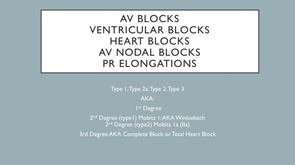
AV Nodal Blocks (heart blocks) • Disturbances of the conduction through the heart, occurring at the AV Node • AV Node – damaged/diseased – delay or total block of impulses at the AV Node • This conduction defect can be seen on the ECG
Here’s the key idea - the signal is either getting through: 1- All the time (but taking a little longer than usual). 2- Some of the time. 3- None of the time. All, some, or none of the time. And see, those are the three kinds, or degrees of heart block: first, second, and third degree. All, some, or none.
Causes • Increased vagal tone (parasympathetic nervous system) • IHD (MI) • Endocarditis • Degeneration (age) • Sclerosis (Aortic) • Cardiac surgery trauma
AV Node • AV nodal conduction time is represented on the ECG as the PR segment. • But - we always measure the PR interval.
First Degree Heart Block (1º) • SA Node – normal • Normal P wave • AV Node conducts more slowly than normal • Prolonged PR Interval • Rest of conduction is normal • Normal QRS
First Degree Heart Block (1º) • PR Interval > 0.2 seconds (5 small sq) • Note – the PR Interval is constant
Significance • Clinical significance • None • Treatment • None • Note – this can progress to 2º or 3º heart block
Second Degree Heart Block (2º) • Mobitz Type I (Wenkebach) • Mobitz Type II • 2 : 1
Second Degree Heart Block (2º)Mobitz Type I(Wenkebach) • Conduction through the AV Node – progressively delayed until a drop beat is seen • Karl Wenkebach
Second Degree Heart Block (2º)Mobitz Type I(Wenkebach) • PR Interval prolongs with each beat until a dropped beat is seen • The PR Interval is NOT constant • After each dropped beat, the PR interval is normal and the cycle starts again
Second Degree Heart Block (2º)Mobitz Type I(Wenkebach) PR PR PR DROPPED BEAT
Significance • Clinical Significance • Slight symptoms eg. Lethargy,Confusion • Treatment • Pacemaker if during day &/or symptoms • No treatment if at night • Note – this can progress to 3º Heart Block
Second Degree Heart Block (2º)Mobitz Type II • Conduction through the AV node is constant. • PR interval is normal and constant • Occasionally a dropped beat is seen
Second Degree Heart Block (2º)Mobitz Type II PR PR DROPPED BEAT PR
Significance • Clinical significance – this is more significant disease • Treatment – pacemaker • Note – this can progress to 3º Heart Block
Second Degree Heart Block (2º) 2 : 1 • Unable to strictly classify as Mobitz Type I or II • Particular type of second degree Heart Block • Ratio 2 P waves : 1 QRS
Significance • Clinical significance – unable to classify as Mobitz type I or II • Will be associated with symptoms, dizziness, lethargy etc. • Treatment – pacemaker • Note – this can deteriorate to 3º Heart Block
Third Degree Heart Block (3º)(Complete) • Complete failure of the AV Node • No impulses from Sinus Node will pass through to the ventricles • Some part if the conducting system will take over as pacemaker of the heart (even a myocardial cell 10-15 bpm)
Third Degree Heart Block (3º)(Complete) • P wave rate – normal • Ventricular rate – slow • Ventricular complex may be broad • Idioventricular rhythm • Complete dissociation between P waves & QRS
Third Degree Heart Block (3º)(Complete) P P P P P QRS QRS
Significance • clinical significance • Symptoms LOC, Confusion, Dizziness, Low BP • Can lead to standstill, VT or VF (stokes Adams) • Treatment - pacemaker
Summary • 1º - prolongation of PR Interval ALL • 2º - Mobitz I – Increasing PR Interval until dropped beat is seen SOME Mobitz II – Constant PR Interval with more P waves to QRS • 2 : 1 – Constant PR Interval with more P waves to QRS • 3º - Complete dissociation between P waves & QRS NONE