THE BASIC SURGICAL SKILLS
THE BASIC SURGICAL SKILLS. Inflammation and wound healing. Vascular response Initial vasoconstriction as a direct response to trauma Exposed subendothelial tissue activates coagulation and complement cascades Platelet adhesion and aggregation causes clot formation

THE BASIC SURGICAL SKILLS
E N D
Presentation Transcript
Inflammation and wound healing • Vascular response • Initial vasoconstriction as a direct response to trauma • Exposed subendothelial tissue activates coagulation and complement cascades • Platelet adhesion and aggregation causes clot formation • Degranulation of platelets releases growth factors and chemotactic factors • Inflammatory response due to histamine and 5HT release produces: • Vasodilatation • Increased capillary permeability • Margination of neutrophils • Cellular response • Migration of neutrophils, macrophages and lymphocytes • Macrophages produce growth factors leading to migration of fibroblast and epithelial cells. • This causes cellular proliferation with three components: • Epithelialisation • Contraction • Fibroplasia
Epithelial barrier important to prevent infection and maintain fluid balance • Achieved by both migration and proliferation of epithelial cells • Migration require presence of granulation tissue • When epithelial cover complete contact inhibition prevents further epithelial growth • Contraction can account for up to 80% reduction in wound size • Contraction due to myofibroblasts in granulation tissue • Fibroplasia due to procollagen production by fibroblasts • Intra and intermolecular bonds form the Collagen fibres with triple helical quaternary structure • Extracellular matrix contains Fibronectin and Glycosaminoaglycans • Regulates collagen synthesis and cellular differentiation • Accompanied by simultaneous angiogenesis • Proliferation is followed by remodeling • Maximum collagen production occurs at 20 days • Maximum wound strength at 3 to 6 months • Initial collagen production disorganised • Remodelling lines it up with stresses in skin • Reduced vascularity and cellularity
Important growth factors • Platelet Derived Growth Factors (PDGF) • Insulin Like Growth Factor (IGF-1) • Epidermal Growth Factor (EGF) • Transforming Growth Factor (TGF) Factors influencing wound healing • Systemic factors • Age and Sex • Nutrition • Vitamin and trace element deficiencies - vitamin C, vitamin A, zinc • Drugs – steroids, chemotherapy, immunosuppression • Systemic disease – diabetes, jaundice, malignancy • Hypoxia • Local Factors • Blood Supply • Infection • Foreign Bodies • Surgical Technique
Scars and contractures • Scar formation • Factors influencing scar formation • Individual genetic make up • Race • Anatomical site • Wound tension • Age • Placement of incision • Surgical technique • To minimise the degree of postoperative scarring: • Incisions should run along Langer's lines • The finest suture possible should be used • Tension should be avoided • Sutures should be removed as soon as possible • Traumatic wounds should be clean and edges excised • Exposure to sunlight should be avoided in the early postoperative period
Problematic scars Contractures • Result if scars shorten • Particularly seen in badly aligned scars not corresponding to Langer's lines • Can reduce joint mobility • May require a z-plasty or skin graft Depressed scars • Result if skin becomes attached to deep tissue • Can be treated by release of normal skin from margins of scar • Scar is then de-epithelialised and skin edges closed over the top Keloid and hypertrophic scars • All scars become red and thickened during the normal healing process • After several months maturation results in flattening of the wound • In some scars collagen formation is excessive • Results in elevated and red scar • If confined to wound = hypertrophic scar • If extends beyond wound into normal tissue = keloid scar • Seen particularly in patients of Afro-Caribbean origin • Particularly affects scars on the presternal and deltoid areas
Surgical technique • Incisions and wound closure • Diathermy, laser • Diathermy • Laser • Sutures and ligature materials • Fundamental skill • Surgical drains
Wound closure 1. Simple interrupted • Insert needle through the skin at right angles including enough subcutaneous tissue to aid in everting the skin edges • Exit through skin of the opposite edge at the same angle including a comparable volume of subcutaneous tissue.
2. Simple continuous • Insert needle as in [1] • Continue suturing without tying ,over and over at fixed intervals and tie at the end. 3. Vertical Mattress
3. Subcuticular continuous • Starting from one end of the incision, insert needle through the dermis taking small bites alternately on one side and then the other • Maintain sutures at same depth from the edge on both sides. • At the end the sutures can be taped to the skin or tied over a dressing.
Diathermy • Diathermy is the use of high frequency electric current to produce heat • Used to either cut or destroy tissue or to produce coagulation • Mains electricity is 50 Hz and produces intense muscle and nerve activation • Electrical frequency used by diathermy is in the range of 300 kHz to 3 MHz • Patients body forms part of the electrical circuit • Current has no effect on muscles
Monopolar diathermy • Electrical plate is placed on patient and acts as indifferent electrode • Current passes between instrument and indifferent electrode • As surface area of instrument is an order of magnitude less than that of the plate • Localised heating is produced at tip of instrument • Minimal heating effect produced at indifferent electrode
Bipolar diathermy • Two electrodes are combined in the instrument (e.g. forceps) • Current passes between tips and not through patient
Effects of diathermy • The effects of diathermy depends on the current intensity and wave-form used • Coagulation • Produced by interrupted pulses of current (50-100 per second) • Square wave-form • Cutting • Produced by continuous current • Sinus wave-form Risk and complications • Can interfere with pacemaker function • Arcing can occur with metal instruments and implants • Superficial burns if use spirit based skin preparation • Diathermy burns under indifferent electrode if plate improperly applied • Channeling effects if used on viscus with narrow pedicle (e.g. penis or testis)
Lasers • Laser = Light Amplification by the Stimulated Emission of Radiation • Laser emissions are: • Collimated - parallel output beam results in little energy loss • Coherent - waves are all in phase resulting in little loss of energy • Monochromic - all of the same wave length • Effects of laser depends on photochemical, photomechanical and photothermal effects • Tissue penetration increases with wavelength • Pulsing of output can reduce thermal damage
Laser safety • Lasers are classified according to the amount of damage they can cause • Class 1 - generally safe • Class 2 - safe within the time of the blink reflex • Class 3 - cause blindness after short exposure from mirrored surfaces • Class 4 - unsafe even with reflection from non-mirrored surfaces • All medical lasers belong to class 4 • Both patients and operators require to wear goggles
Risks associated with lasers • To patient • Excessive burning • Scar formation • Visceral perforation • To the operator • Accidental skin exposure • Corneal or retinal burns
Choosing the right suture material • Absorbable and Non-absorbable • Monofilament and Multifilament • Natural and synthetic • Suture diameter and strength
Choice of a suture Choice of suture depends on: • Properties of suture material • Absorption rate • Handling characteristics and knotting properties • Size of suture • Type of needle
Suture characteristics • Suture materials vary in their physical characteristics • Monofilament sutures (e.g. polypropylene) are smooth • The slide well in tissues but if handles inappropriately they can fracture • Multifilament sutures (e.g. polyglactin) are braided • They have a greater surface area • They are easier to handle and knot well • Some suture materials have a 'memory' (e.g. polypropylene) • Return to former shape when tension is removed
1. Catgut • Made from the submucosa of sheep gastrointestinal tract • Broken down within about a week • Chromic acid delays hydrolysis • Even so it is destroyed before many wounds have healed 2. Silk • Strong and handles well but induces strong tissue reaction • Capillarity encourages infection causing suture sinuses and abscesses 3. Vicryl • Tensile strength • 65% @ 14 days • 40% @ 21 days • 10% @ 35 days • Absorption complete by 70 days 4. Polydioxone (PDS) • Tensile strength • 70% @ 14 days • 50% @ 28 days • 14% @ 56 days • Absorption complete by 180 days
Absorbable suture are broken down by either: • Proteolysis (e.g. Catgut) • Hydrolysis (e.g. Vicryl, Dexon)
Suture sizes • Sutures are sized by the USP (United States Pharmacopoeia) scale • The available sizes and diameters are: • 6-0 = 0.07 mm • 5-0 = 0.10 mm • 4-0 = 0.15 mm • 3-0 = 0.20 mm • 2-0 = 0.30 mm • 0 = 0.35 mm • 1 = 0.40 mm • 2 = 0.5 mm
Surgical drains • Drains are inserted to: • Evacuate establish collections of pus, blood or other fluids (e.g. lymph) • Drain potential collections • Their use is contentious • Arguments for their use include: • Drainage of fluid removes potential sources of infection • Drains guard against further fluid collections • May allow the early detection of anastomotic leaks or haemorrhage • Leave a tract for potential collections to drain following removal • Arguments against their use include: • Presence of a drain increases the risk of infection • Damage may be caused by mechanical pressure or suction • Drains may induce an anastomotic leak • Most drains abdominal drains infective within 24 hours
Types of drains • Drains can be: • Open or closed • Active or passive • Drains are often made from inert silastic material • They induce minimal tissue reaction • Red rubber drains induce an intense tissue reaction allowing a tract to form • In some situations this may be useful (e.g. biliary t-tube)
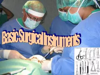
Fundamental Skills Handling of Instruments, Knots by hand and instruments Skin Suture Tissue Debridement Tendon Suture
TISSUE DEBRIDEMENT • This is the most essential step in the management of the contaminated or dirty wound. • Thoroughly wash the wound with copious amounts of normal saline under pressure using either a large syringe , rubber bulb or a pulsatile lavage system. • Remove large particles of dirt with a toothed forceps. • Sharply excise [ using a No. 10 blade scalpel or sharp scissors] all slough, devitalised tissues and grossly contaminated tissues. • Wash again.
TENDON SUTURING Instruments • Hypodermic needles • Small curved hemostat • Fine non-toothed forceps • Needle holder • 3-0 and 5-0 prolene sutures • Suture cutting scissors
Preparation • Match correct tendon ends using the position of the tendons & the corresponding shapes of the cut ends. • Fix tendon with a needle and steady one end with a needle. • Do not grasp tendons with any instrument as this will promote adhesions. Core Suture • Insert the 3-0 prolene suture through the cut end of the tendon and exit through' the side at least 1cm away from the end • Continue transversely through' the tendon and reinsert suture from the opposite side , this time to exit through' the cut end adjacent to the entry point • Repeat the above for the distal end of the tendon • Tie the ends [1 double throw and at least 4 single throws] such that the knot is buried in between the cut ends Epitendinous suture • Using 5-0 prolene, run a simple running suture through the epitenons of the cut edges circumferentially taking care not to injure the core suture • Tie the knot at the end
HAND SUTURED BOWEL ANASTOMOSIS BY SEROSUBMUCOSAL TECHNIQUE 1.Types • Interrupted serosubmucosal suture ( the "gold standard" for intestinal anastomosis). • Continuous serosubmucosal suture. 2.The Basis • Faster and sounder healing when compared to the traditional two layer anastomosis. 3. Advantages • Accurate tissue apposition. • Incorporates submucosa - the strongest layer. • Minimises damage to submucosal vascular plexus. • Lesser tissue strangulation. • Lesser reduction in lumen size. • Interrupted suture accommodates luminal discrepancies up to 50% without resorting to an antimesenteric slit. No 'purse string' effect even with continuous suture. • Minimises risk of implantation of neoplastic cells. • Appropriate for both upper and lower GI tract anastomosis. Continuous suture more useful in upper GI anastomoses (biliary, pancreatic). • Appropriate for both accessible and inaccessible sites. • Continuous suture is faster than interrupted suture.
Factors influencing anastomotic healing • Anastomotic technique is required to maintain apposition until collagen is laid down • Anastomoses show serosal healing and require: • Maintenance of apposition • Good blood supply • Tension free • Anastomotic leak or failure may occur if: • Distal obstruction • Peri-anastomotic sepsis • Per-anastomotic haematoma • Hypotension • Hypoxia • Jaundice • Corticosteroids • Uraemia