Download
1 / 19
240 likes | 1.17k Vues
Advanced Medicinal Chemistry. Lecture 3:. Molecular Interactions and Drug Potency. Barrie Martin AstraZeneca R&D Charnwood. Agonists: Measure % Response vs Agonist concentration EC 50 - The agonist concentration that causes 50% of the maximum response. pEC 50 = - log 10 (EC 50 ).

E N D
Advanced MedicinalChemistry Lecture 3: Molecular Interactions and Drug Potency Barrie Martin AstraZeneca R&D Charnwood
Agonists: Measure % Response vs Agonist concentration EC50 - The agonist concentration that causes 50% of the maximum response. pEC50 = - log10(EC50) Dose-Response Curves 100 Enzyme Inhibitors (competitive): Measure inhibition at differing concentrations of ‘drug’. % Response % Inhibition 50 IC50 - The inhibitor concentration that causes a 50% reduction in intrinsic enzyme activity IC50=85nM EC50=85nM 0 10nM 30nM 100nM 300nM 1mM pIC50 = - log10(IC50) IC50 1M = pIC50 6.0 IC50 1nM = pIC50 9.0 [Inhibitor] [Agonist] Antagonists: Situation more complex. Antagonists displace the agonist dose-response curve rightwards – most accurate measure of potency (pA2) requires measurement of agonist binding at multiple concentrations of antagonist For a drug, typically target affinity values of pIC50 8 (<10 nM concentration)
iNOS - An AZ Charnwood Discovery Project Active Site, Haem & Inhibitor Nitric Oxide Synthases – catalyse production of NO from arginine in the body – implicated in inflammatory conditions e.g. rheumatoid arthritis AZ10896372 pIC50 7.5 A potent, selective iNOS inhibitor
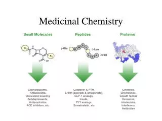
How Do Drugs Bind to Enzymes & Receptors? Drugs bind to particular sites on enzymes and receptors. In the case of an enzyme, this will often be theactive site. Receptors havebinding pocketsformed between transmembrane helixes where drugs usually bind (not always the agonist’s binding site). GLUE PHEF • These sites are comprised of a variety of amino acid residues which give rise to a specific 3-D shape and molecular features: • Charges: CO2- , NH3+, =NH-+ • Polar groups: OH, C=O, CONH • Hydrophobic groups: Ph, Alkyl, SMe SERS • In enzymes, reaction centres are also present: • Asp-His-Ser in esterases • SH in some proteases • Metal ions (CYP-450, iNOS). • Small molecules bind to these pockets by a combination of: • Shape complementarity • Energetically favourable interactions Haem group – iNOS, CYP-450
Shape Complementarity iNOS Enzyme Inhibitor AZ10896372 Arginine H2 Receptor Antagonist H H N N Cimetidine Histamine S H N N C N N H 2 H N N • The drug must fit into theBinding Siteand shape complementarity is an important feature of a drug molecule. Competitive enzyme inhibitors often bear a resemblance to the substrate, as they bind to the sameActive Site. This is also true forsomereceptor antagonists,but not all. • The strength of an interaction depends on the complementarity of the physico-chemical properties of atoms that bind, i.e. protein surface and ligand structure. • The ‘Binding Sites’ are not totally rigid. The side chains of the amino acids that make up the pocket have some mobility. A variety of related structures can thus be accommodated by movements that change the shape of the active site. This is known as the‘Induced Fit Hypothesis’.
Drug-Protein Binding Energies For a binding Equilibrium between a Protein & a Drug K [Protein] + [Drug] [P:D] DG Drug Protein Protein Drug Gibbs Free Energy Changes K = [P:D] [P] x [D] DG=-RTlnK and DG=DH-TDS Both Enthalpy (DH) and Entropy (DS) changes affect binding strength
Drug-Protein Interactions Bond Example kJ/mol Van der Waal Xe…Xe, alkyl groups 2 Hydrophobic Ph…Ph (p-stacking)5 Dipole - Dipole C=O…HN-R (d+/d-)...(d+/d-) 5 Hydrogen H2O…H2O (X-H) …(Y-R) 35 Ion - Dipole F-…H2O (+/-ve)…(d+/d-) 170 Ion - Ion H+…Cl- (+ve)…(-ve) 450 Covalent C-O 350 NB. When a drug moves from the aqueous medium into the ‘Binding Site’ it has to break H-Bonds with water, de-solvate etc. These processes require energy, so the netenergy available for binding is only a fraction of the above bond energies.
Electrostatic Interactions AZ-10896372 iNOS Inhibitor Neuraminidase Inhibitor (Antiviral GSK) O H H F N N H N N H A R G N + O H O N - H H N N O + H O H O O F N O H H H R O GLU R • These result from the attraction between molecules bearing opposite • electronic charges. • Strong ionic interactions can contribute very strongly to binding. • Proteins contain bothCO2-andNH3+residues and these may be present • at the binding site to interact with oppositely charged groups on the drug. • The energies involved in a‘salt bridge’can be in the order of>30 kJ/mol • This can lead to increase in observed binding of>106 fold
Hydrogen Bonding Interactions A hydrogen bond results when a hydrogen is shared between two electronegative atoms TheDonorprovides the H, while theAcceptorprovides an electron pair e.g. R-O-H…..O=C D-X-H….Y-A O H O H O N N O H O H O H O R O H O R O H H G L U O O H H N N Neuraminidase Inhibitor Charge re-inforced H-Bond N N N + H F N H H AZ10896372 - iNOS complex Amide to Tyrosine H-Bond
Hydrophobic Interactions • Drugs, in general, are hydrophobic molecules • The ‘Binding Sites’ of proteins are also hydrophobic in character • Thus a mutual attraction can result (like attracts like). • What drives this attraction? • Enthalpy gains may result from van der Waals bonding: • Between Alkyl, Aryl, Halogen groups • p-p Stacking is an important type of this • Entropy gains are achieved when water molecules are displaced • from ‘active site’, and return to a more random (high S) state. • Each -(CH2)- group can contribute >1 kJ/mol towards binding • Each -Ph ring can contribute >2 kJ/mol towards binding • These effects are additive and henceHydrophobic Bonding • can make a very high contribution to binding
Water molecules are in a highly disordered state. Each molecule maximises H-Bonds to other molecules of water. When a hydrophobic drug is placed into water, the structure of the water around the drug is more ordered. This allows the H2O-H2O H-bonds to be maintained. This leads to lowerentropy and is not favoured. Hydrophobic Bonding : D Entropy
D E E D • Hydrophobic interaction between protein and drug is favoured byentropy gains: • Bulk water returns to less ordered state • Water molecules may be expelled from being bound in active site. • In additionenthalpygains due to new bonds may also be favourable (e.g. van der Waals interactions) Hydrophobic Bonding : D Entropy
Probing Hydrophobicity in Drug Discovery New iNOS lead identified: R =Me, small lipophilic substituent iNOS pIC50 7.8 Aim: Probe lipophilic pocket – what else could we put there? How would we make it?
Effect of Hydrophobicity on Activity 8.6 8.4 8.2 iNOS_pIC50 8 7.8 Too big to fit in pocket optimally (Shape complementarity) 7.6 1 1.2 1.4 1.6 1.8 2 2.2 2.4 2.6 cLogP Binding into Lipophilic pocket of iNOS R cLogP IC50 mM Me 1.13 0.016 Et 1.66 0.009 CF3 1.75 0.008 Thiophene 2.02 0.003 Phenyl 2.34 0.015 2-Me-Thiophene 2.48 0.026
Bioisosteres Isostere: Similarities in physicochemical props. of atoms/groups/molecules with similar electronic structures (no. and arrangement of electrons in outermost shell). Often observed with groups in the same periodic table column (Cl Br, C Si). Grimm – Hydride Displacement Law (1925) - Replacement of chemical groups by shifting one column to the right & adding H. Bioisostere: Simplest definition - any group replacement which improves the molecule in some way Two different interchangeable functionalities which retain biological activity. Bioisosteric replacements can offer improvements both in potency and other properties (e.g. metabolic stability, absorption) -CH2 & bioisosteres Carboxylic acid & bioisosteres amide & bioisosteres
Invisible Bioisosteres EGF-R 2.2 nM EGF-R 7.5 nM H-bonds can be directly to protein or via water molecules
Optimising Potency How might we improve potency further from this compound? Develop understanding of which molecular features are important for activity – remove substituents. Look at incorporating new groups for additional potency e.g. through lipophilic interactions, hydrogen bonds etc. Functional group bioisosteres. Use available structural information – e.g. crystal structures of compound bound to enzyme. Use of modelling to design/evaluate new targets. Develop and test hypotheses. Identify good disconnections/robust chemistry to allow rapid synthesis of multiple analogues – build up information. pIC50 7.5 N.B. Potency is one of many properties that needs to be optimised in drug discovery - need to consider absorption, metabolism, selectivity etc.