Download
1 / 27
340 likes | 1.14k Vues
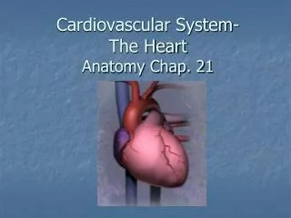
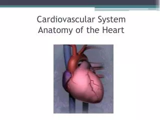
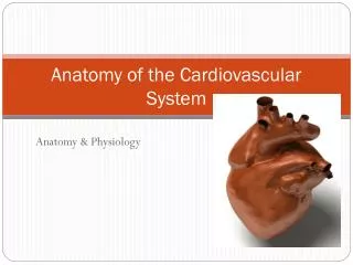
Anatomy of the Cardiovascular System. Anatomy & Physiology. Heart song. http://www.youtube.com/watch?v=q0s-1MC1hcE. Location of the heart. In mediastinum behind the sternum between 2 nd & 6 th ribs. Shifted to left. Posteriorly between 5 th to 8 th thoracic vertebrae

E N D
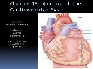
Anatomy of the Cardiovascular System Anatomy & Physiology
Heart song • http://www.youtube.com/watch?v=q0s-1MC1hcE
Location of the heart • In mediastinum behind the sternum between 2nd & 6th ribs. Shifted to left. • Posteriorly between 5th to 8th thoracic vertebrae • Apex (lowest point) lies on diaphragm
Pericardium • Sac covering the heart • Fibrous pericardium – tough, inelastic • Serous pericardium- 2 layers • Parietal layer- lines fibrous pericardium • Visceral layer (epicardium)-adheres to outside of heart; space between parietal & visceral layer contain small amount of fluid
Function of pericardium • Provides protection against friction
Layers of the heart • Epicardium (also the serous pericardium) • Myocardium-thick contractile layer of muscle cells • Endocardium-cover trabeculae (muscular projections); specialized folds of endocardium make up the major valves of heart
Chambers of heart • Atria: upper chambers • Ventricles: lower chambers • Chambers are separated by an extension of heart wall, septum
Atria • Receive blood from veins • Send blood to ventricles • Thin myocardial wall • Auricle: earlike flap protruding from each atrium
Ventricles • Receive blood from the atria • Pump blood out of heart into arteries • Thicker myocardium • Myocardium of left ventricle thicker than right ventricle
Heart valves • Permit flow of blood in one direction • Atrioventricular valves • Semilunar valves
Atrioventricular valves • Made of flaps of endocardium • Flaps anchored to papillary muscle by chordae tendineae • Rt AV valve: tricuspid valve • Lt AV valve: mitral (bicuspid) valve
Semilunar valves • Consists of half moon shaped flaps • Pulmonary semilunar valve: at the entrance to the pulmonary artery • Aortic semilunar valve: at the entrance to aorta
Heart video • http://www.youtube.com/watch?v=DAXa4eR1s0M
Blood supply to heart • Blood supplied thru the right & left coronary arteries • Coronary arteries are first branches off aorta • After blood has passed thru capillary beds, enters cardiac veins then thru common channel, coronary sinus, then into rt atrium
Conduction system of heart • Sinoatrial node (pacemaker): in rt atrium near opening of superior vena cava • Atrioventricular node: in rt atrium along lower part of interatrial septum • Atrioventricular bundle: originate in AV node & extend by 2 branches thru the 2 sides of the interventricular septum • Purkinje fibers: extension of AV bundle out to the lateral walls of the ventricles & papillary muscles
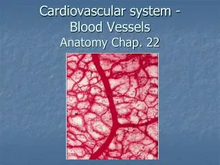
Blood vessels • Arteries: carry blood away from heart • Arterioles: small arteries • Veins: Carry blood toward heart • Venules: small veins • Capillaries: microscopic blood vessels that carry blood from arterioles to venules
3 layers of blood vessels • Tunica adventitia: outer layer, fibrous connective tissue • Tunica media: layer of smooth muscle & elastic connective tissue • Tunica intima: made of smooth endothelium
Functions of capillaries • Transport materials to and from cells • Capillaries so numerous & so small that blood flows at its slowest rate in capillaries
Functions of arteries • Function as “distributors” • Arterioles also function as resistance vessels • Smooth muscle cells on arterioles act as precapillary sphincters where a capillary originates
Functions of veins • Return blood to heart • Act as reservoir vessels • Ability to stretch by veins called capacitance thus veins are capacitance vessels
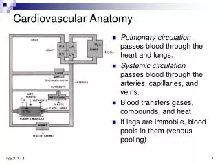
Circulatory routes • Systemic circulation: blood flow from heart to all parts of body (except lungs) • Pulmonary circulation: blood flow from heart to lungs
Arteries • End-arteries: most of the arteries, they diverge into capillaries • A few arteries open into branches of other arteries; this communication is an arterial anastomosis • Incidence of arterial anastomosis increases as distance from the heart increases
Aorta • Major artery that serves as trunk of the entire systemic arterial system • First few cm conducts blood up away from lt ventricle-ascending aorta • Then turns 180 degrees-aortic arch • Then downward from arch-descending aorta
Veins • Large veins that return blood to heart in systemic circulation are superior & inferior vena cava