Download
1 / 36
370 likes | 402 Vues
Learn about key muscles surrounding hip and knee joints, their movements, and functions in leg and thigh actions.

E N D
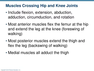
Muscles Crossing Hip and Knee Joints • Include flexion, extension, abduction, adduction, circumduction, and rotation • Most anterior muscles flex the femur at the hip and extend the leg at the knee (foreswing of walking) • Most posterior muscles extend the thigh and flex the leg (backswing of walking) • Medial muscles all adduct the thigh
12th thoracic vertebra 12th rib Quadratus lumborum Psoas minor Iliac crest Psoas major Iliopsoas Iliacus 5th lumbar vertebra Anterior superior iliac spine Tensor fasciae latae Pectineus Adductor longus Sartorius Gracilis Quadriceps femoris Adductor magnus • Rectus femoris • Vastus lateralis • Vastus medialis Tendon of quadriceps femoris Patella Patellar ligament (a) Figure 10.19a
Movements of the Thigh • Thigh extensors • Hamstring muscles (prime movers of extension) • Biceps femoris • Semitendinosus • Semimembranosus • Gluteus maximus (prime mover during forceful extension)
Gluteus medius Gluteus maximus Adductor magnus Gracilis Iliotibial tract Long head Biceps femoris Short head Hamstrings Semitendinosus Semimembranosus (a) Figure 10.20a
Movements of the Thigh • Adductors (also medially rotate thigh) • Adductor magnus • Adductor longus • Adductor brevis • Gracilis
Pectineus (cut) Adductor brevis Adductor magnus Adductor longus Femur O = origin I = insertion (b) Figure 10.19b
Movements of the Thigh • Abductors • Gluteus maximus (also laterally rotates thigh) • Gluteus medius (also medially rotates thigh) • Gluteus minimus (also medially rotates thigh)
Gluteus medius (cut) Gluteus minimus Superior gemellus Piriformis Obturator internus Obturator externus Quadratus femoris Inferior gemellus Gluteus maximus (cut) (c) Figure 10.20c
Muscles of the Thigh that Move the Knee Joint • Quadriceps femoris—sole extensor of the knee • Hamstring muscles—flex the knee, and are antagonists to the quadriceps femoris
12th thoracic vertebra 12th rib Quadratus lumborum Psoas minor Iliac crest Psoas major Iliopsoas Iliacus 5th lumbar vertebra Anterior superior iliac spine Tensor fasciae latae Pectineus Adductor longus Sartorius Gracilis Quadriceps femoris Adductor magnus • Rectus femoris • Vastus lateralis • Vastus medialis Tendon of quadriceps femoris Patella Patellar ligament (a) Figure 10.19a
Gluteus medius Gluteus maximus Adductor magnus Gracilis Iliotibial tract Long head Biceps femoris Short head Hamstrings Semitendinosus Semimembranosus (a) Figure 10.20a
Muscles of the Leg: Movements • Various leg muscles produce the following movements • Ankle—dorsiflexion and plantar flexion • Intertarsal joints—inversion and eversion of the foot • Toes—flexion and extension
Muscles of the Anterior Compartment of the Leg • Primary toe extensors and ankle dorsiflexors • Tibialis anterior • Extensor digitorum longus
Fibularis longus Gastrocnemius Tibia Tibialis anterior Extensor digitorum longus Soleus Extensor hallucis longus Fibularis tertius Superior and inferior extensor retinacula Extensor hallucis brevis Extensor digitorum brevis (a) Figure 10.21a
Patella Head of fibula Gastrocnemius Soleus Fibularis longus Extensor digitorum longus Tibialis anterior Extensor hallucis longus Fibularis tertius Fibularis brevis Superior and inferior extensor retinacula Flexor hallucis longus Extensor hallucis brevis Fibular retinaculum Extensor digitorum brevis Lateral malleolus (a) 5th metatarsal Figure 10.22a
Muscles of the Posterior Compartment of the Leg • Flexors of the foot and the toes • Gastrocnemius • Soleus
Plantaris Medial head Gastrocnemius Lateral head Tendon of gastrocnemius Calcaneal tendon Medial malleolus Lateral malleolus Calcaneus (a) Superficial view of the posterior leg. Figure 10.23a
Hamstrings Adductors Vastus lateralis Femur Posterior compartment of thigh (flexes leg and extends thigh); innervation: tibial nerve (portion of sciatic nerve) Vastus intermedius Rectus femoris Vastus medialis (a) Posterior compartment muscles Anterior compartment muscle Medial compartment muscles of thigh and lateral compartment muscles of leg Medial compartment (adducts thigh); innervation: obturator nerve Anterior compartment (extends leg); innervated by femoral nerve (a) Muscles of the thigh Figure 10.25a
Posterior compartment muscles Triceps surae Anterior compartment muscle Fibula Fibularis muscles Medial compartment muscles of thigh and lateral compartment muscles of leg Posterior compartment of leg (plantar flexes foot, flexes toes); innervated by tibial nerve (b) Tibialis anterior Tibia Lateral compartment of leg (plantar flexes and everts foot); innervation: superficial fibular nerve Anterior compartment of leg (dorsiflexes foot, extends toes); innervated by deep fibular nerve (b) Muscles of the leg Figure 10.25b
Fibularis longus Gastrocnemius Tibia Tibialis anterior Extensor digitorum longus Soleus Extensor hallucis longus Fibularis tertius Superior and inferior extensor retinacula Extensor hallucis brevis Extensor digitorum brevis (a) Figure 10.21a
Question: Which kite shaped muscle pair stabilizes, raises, retracts, and rotates the scapula? Answer: Trapesius
Which fascicle patter is represented by letter (b)? Which fascicle pattern is represented by letter (e)? Name the fascicle pattern represented by letter (d) Which letter would represent a fusiform fascicle pattern?
Which of these levers is a speed lever? Which of these levers is at a mechanical disadvantage? Explain the relationship between the load, fulcrum and effort when classifying a lever as a mechanical advantage compared to a disadvantage.
Point to platysma Point to and name the muscle that is the antagonist to depressor labii inferioris Identify and name the prime mover for chewing Point to and name the muscle used for whistling and nursing infants Identify and name the muscle that is a synergist for jaw closure/chewing
Locate the linea alba Locate serratus anterior Name and locate the prime mover for spinal flexion Locate the transverse abdominals
Locate and identify the muscle in this image that is part of the rotator cuff Locate pectoralis major Locate and identify the muscles inferior to pectoralis minor Locate and identify the muscle responsible for flexion and lateral rotation of the neck Locate and identify the prime mover for arm abdcuction
Locate and identify the muscle responsible for arm adduction Locate teres minor Locate rhomboid major Locate trapezious
Locate gracillis Locate sartorious Which group of muscles does gracilis fit into Locate vastus lateralis List the muscles included in the quadricep group and give the main function Which muscle of the quadricep group is not visible in this image?