
Innovations in FACS Analyzers: Enhanced Optical Design Reduces Footprint and Increases Functionality
In recent years, advancements in optical design have significantly improved the functionality and compactness of FACS analyzers. A study comparing the Gallios and SORP LSR II flow cytometers demonstrated that the Gallios provided 89% event recovery compared to only 11% for the LSR II when analyzing pre-enriched mouse lymphocytes. The enhanced optical pathway and scatter properties of the Gallios led to superior separation, resolution, and ease of population identification. These improvements reflect a necessary shift in flow cytometry instrumentation to meet the demands of the growing research community.

Innovations in FACS Analyzers: Enhanced Optical Design Reduces Footprint and Increases Functionality
E N D
Presentation Transcript
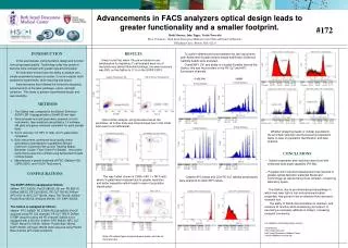
Advancements in FACS analyzers optical design leads to greater functionality and a smaller footprint.  #172 Heidi Mariani, John Tigges, Vasilis Toxavidis Flow Cytometry, Beth Israel Deaconess Medical Center/ Harvard Stem Cell Institute 3 Blackfan Circle, Boston, MA, 02215 RESULTS It was found that, when 10k pre-enriched mouse lymphocytes for regulatory T cell analysis were run at manufacturers default threshold settings, the event recovery was 89% on the Gallios to 11% on the SORP LSR II. INTRODUCTION In the past decade, instrumentation design and function have progressed greatly. Technology today has grown to become more compact with greater optical functionality. An instrument should have the ability to analyze very simple experiments based on scatter, to more complex multi-parametric experiments, while requiring less space. Instrumentation has followed this format by designing advancements in the laser pathways, optics, and light collection. This leads to greater experimental design and reproducibility. • Greater separation and resolution was found with enhanced wide angle capability (FS W2). • Progress with instrument development has resulted in greater optical detection (patented Boulevard Technology) as well as being more compact, conserving laboratory space. The Gallios, due to an enhanced optical pathway in which less laser light is lost and enhanced scatter properties, has proven to be an effective flow cytometric research tool. The ability of FACS instrumentation to maintain, and enhance its function while downsizing its footprint, is becoming a necessary attribute in today’s increasing research community. To confirm differences found between the two instruments, both Spherotech 8 peak rainbow beads and Becton Dickinson Calibrite beads were analyzed. Overall MFI, CV, and ability to visualize 8 peaks favored the Gallios; this was most evident in the PE-Cy7 and APC fluorescent channels. GALLIOS LSRII METHODS The Gallios was compared to the Becton Dickinson SORP LSR II equipped with a 50mW 561nm laser. Various beads and cell types were acquired on both instruments, then analyzed using Kaluza 1.0 software. (All plots and gates remained consistent for each sample type). Event recovery, CV, MFI, % total, and % gated were compared. Both instruments underwent initial quality check according to manufacturer’s guidelines (Becton Dickinson- Cytometer Set-up and Tracking Beads; Beckman Coulter- Flow Check Pro Beads). Instrument performance was then verified using Spherotech 8 peak rainbow beads. Manufacturer’s preset threshold off FSC (Gallios=100; LSRII=5000), and 10,000 Total events. Upon further analysis, using fluorescence as the parameter, all further data was compromised due to this initial total event count differential. • Whether analyzing beads or cellular populations, the enriched resolution and fluorescence separation leads to ease of population identification and data analysis. CONCLUSIONS CONFIGURATIONS This was further shown in CD56+ NK1.1+ NKT cells; where % gated was increased due to greater resolution and better separation which leads to ease of population identification. Calibrite APC beads and CD4 PE-Cy7 labeled lymphocytes were analyzed to obtain MFI values. The SORP LSR II is equipped as follows: 488nm- FITC 530/30, PerCP 685/35; 561nm- PE 585/15, DsRed 585/15, PE Cy5 660/20, PE Cy7 780/60; 640nm- APC 670/14, APC Cy7 780/60, Alexa 700 730/45; 405nm- Pacific Blue 450/50, AmCyan 560/40; UV- DAPI 450/50. The Gallios is equipped as follows: 488nm- FITC 525BP, PE 575BP, PECy5 695/30 (PerCPacquired using PE Cy5 channel), PE Cy7 755LP, DsRed 575BP (acquired using the PE channel; Gallios is not equipped with a 561nm); 633nm- APC 660/20, APC Cy7 755LP, Alexa700 725/20; 405nm- Pacific Blue 450/40, DAPI 450/40, AmCyan 450/40 (both acquired using Pacific Blue channel with violet excitation). For additional information please contact: Heidi Mariani Flow Cytometry Core Beth Israel Deaconess Medical Center hmariani@bidmc.harvard.edu Note: All sample types compared were same, and ran on the same day.


