Download
1 / 1
10 likes | 131 Vues
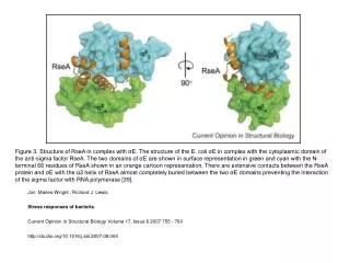
This figure illustrates the structure of E. coli σE in complex with the anti-sigma factor RseA. The complex reveals extensive interactions, with the domains of σE depicted in green and cyan, while the N-terminal 66 residues of RseA are shown in an orange cartoon representation. The α3 helix of RseA is notably buried between the two domains of σE, effectively preventing σE from interacting with RNA polymerase. Such structural insights contribute to our understanding of bacterial stress responses.

E N D
Figure 3. Structure of RseA in complex with σE. The structure of the E. coli σE in complex with the cytoplasmic domain of the anti-sigma factor RseA. The two domains of σE are shown in surface representation in green and cyan with the N-terminal 66 residues of RseA shown in an orange cartoon representation. There are extensive contacts between the RseA protein and σE with the α3 helix of RseA almost completely buried between the two σE domains preventing the interaction of the sigma factor with RNA polymerase [39]. Jon Marles-Wright , Richard J Lewis Stress responses of bacteria Current Opinion in Structural Biology Volume 17, Issue 6 2007 755 - 760 http://dx.doi.org/10.1016/j.sbi.2007.08.004