Download
1 / 26
260 likes | 461 Vues
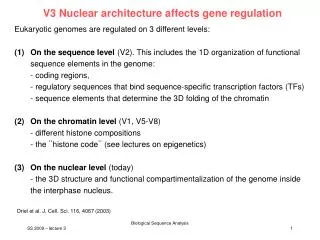
V3 regulation of imprinted genes. Review of lecture V2 ... What does the differential methylation of CpG islands mean? - What do the two models describe? - How did the authors arrive at the two models? - How could one distinguish between these two models?. Outline.

E N D
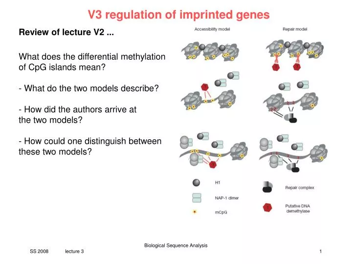
V3 regulation of imprinted genes Review of lecture V2 ... What does the differential methylation of CpG islands mean? - What do the two models describe? - How did the authors arrive at the two models? - How could one distinguish between these two models? Biological Sequence Analysis
Outline • what is genomic imprinting? • networks of imprinted genes • imprinting mechanisms • protein-DNA interaction hypotheses • detecting motifs in DNA sequences • evolutionary conserved regions • protein binding sites • gene regulation modules • "imprinting motifs" • ... • Alu sequences • KCNQ1 Biological Sequence Analysis
Genomic Imprinting • monoallelic expression of a gene depending on its parental origin • in mammals about 70 known imprinted genes (human, mouse), also found in insects and in flowering plants • estimated: 1 - 2% of all genes = 300 - 600 • imprinted genes are often organized in clusters with "imprinting centers" Igf2 H19 H19 Igf2 H19 Igf2 paternal gene copy maternal gene copy Igf2: coding insulin-like growth factor protein H19: untranslated RNA Biological Sequence Analysis
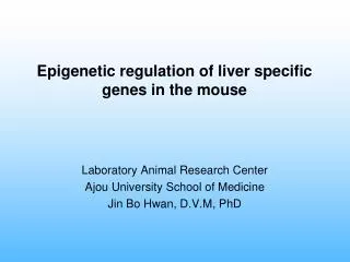
Imprinted genes Imprinted genes of the mouse are distributed unevenly throughout the genome. About half of the known ones are located on Chromosome 7, clustered into at least five distinct imprinted domains. red: maternally expressed genes blue: paternally expressed genes PLOS Genet. 2, e147 (2006) Biological Sequence Analysis
Imprinting Mechanisms • methylation of Cytosine in CpG: differentially methylated regions (DMRs) • altered chromatin structure • binding of proteins (transcription factors, silencers) depending on methylation status • setting the imprint • hypothesis: male specific and female germ line specific proteins recognize different patterns and set different imprints in sperm and egg • how these imprint markers might find their targets: • tandem repeats • sequence not (well) conserved – like many DMRs – • are enriched in the CpG islands of imprinted genes • special DNA structure • sequence patterns (germ line specific protein/transcription factor binding sites): evolutionary conserved AGAACCGCGGCGAGAGGCC AGAACCGCGCCGAAGAACC ACAACCGCGCCGAAGAACC AGAACCGCGCCGAAAAGCC Biological Sequence Analysis
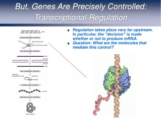
Regulatory models at imprinted loci (A) The enhancer–blocker model (also known as the boundary model) is well studied at the Igf2/H19 locus and consists of an imprinting control region (ICR) located between a pair of reciprocally expressed genes that controls access to shared enhancer elements. On the paternal allele, the differentially methylated domain (DMD) acquires methylation (black circles) during spermatogenesis, which leads to repression of the H19 promoter. The hypomethylated maternal DMD acts as an insulator element, mediated through binding sites for the methylation-sensitive boundary factor CTCF (shaded ellipse). When CTCF is bound, Igf2 promoter access to the enhancers (E) distal to H19 is blocked. Blue boxes : paternally expressed alleles, red boxes : maternally expressed alleles, black boxes : silenced alleles, grey boxes : nonimprinted genes. Arrows on boxes indicate transcriptional orientation. PLOS Genet. 2, e147 (2006) Biological Sequence Analysis
Protein Interactions and Chromatin Loops • reading the imprint: candidate "imprinting transcription factors" CTCF, YY1 • chromatin loop model • DMRs interact via proteins • mediates interaction with the enhancers H19 Igf2 • Murrell et al. (2004) Nature Genet. 36: 889 • maternal chromosome: DMR1 and DMR unmethylated, CTFC bound H19 is expressed (interaction with the enhancers), Igf2 is silenced • paternal chromosome: DMR and DMR2 methylated, no CTCF binding Igf2 in contact with enhancers, active; H19 silenced Biological Sequence Analysis
Regulatory models at imprinted loci (B) At the Igf2r locus on Chromosome 17, the paternally expressed, noncoding RNA Air acts to induce bidirectional cis-mediated silencing (black curved lines) on neighbouring protein-coding genes (maternally expressed Igf2r, Slc22a3, and Slc22a2). The grey ellipses are the intronic imprint control elements that are maternally methylated (black circles) and contain the promoter of the Air RNA. PLOS Genet. 2, e147 (2006) Biological Sequence Analysis
Regulatory models at imprinted loci (C) At microimprinted domains, oocyte-derived methylation in the promoter region of a protein-coding gene is likely to be the primary epigenetic mark leading to monoallelic silencing. With the exception of the U2af1-rs1 locus, the multiexonic genes within which the paternally expressed transcripts are embedded, escape imprinting. The paternally expressed Nap1l5 is situated within intron 22 of Herc3, which is expressed from both alleles. PLOS Genet. 2, e147 (2006) Biological Sequence Analysis
Evolution of imprinted loci Blue: paternally derived alleles, red: maternally derived alleles, Yellow: transposed sequence. Black lollipops: methylated CpGs, light blue dome: a trans-acting factor. Asterisk: gene duplicate. (A) Random molecular events or mutations in the germ-cell lineage generate alleles that undergo differential methylation when passing through the male and female germ line, which can confer either (B) negative or (C) positive fitness. PLOS Genet. 2, e147 (2006) Biological Sequence Analysis
Functions of Imprinted Genes • imprinting disorders generally cause diseases • over- or underexpression of the corresponding gene products • control cell proliferation • growth factors • tumor suppressors • embryonic development ("giant baby") • important for brain development • (adult) behavior • imprinted genes are often transcription factors • regulation of other genes Biological Sequence Analysis
Are the imprinted genes alone? Protein-Protein Interactions of Imprinted Genes • Source: Unified Human Interactome (http://mdc-berlin.de/unihi) Biological Sequence Analysis
Are the imprinted genes alone?Coexpression Network of Imprinted Genes http://symatlas.gnf.org Varrault et al. (2006) Dev. Cell 11: 711 Zac1 (Plagl1) is a transcription factor -> also regulatory networks! Arima et al. (2005) NAR 33: 2650 Biological Sequence Analysis
Imprinted genes and repetitive elements Imprinted genes show depletion of short interspersed transposable elements (SINEs) and an enrichment of long interspersed nuclear element 1 (LINE-1) repeats. Biological Sequence Analysis
Alu sequence An Alu sequence is a short stretch 300 bp of DNA originally characterized by the action of the Alu restriction endonuclease that was isolated from Arthrobacter luteus. They are therefore classified as short interspersed nuclear elements (SINEs) and are the most abundant mobile elements in the human genome. There are over one million Alu sequences of different kinds interspersed throughout the human and other primate genomes, and probably make up about 10% of the whole genome. Less than 0.5% are polymorphic. Alu sequences are derived from the small cytoplasmic 7SL RNA, a component of the signal recognition particle. The recognition sequence of the Alu endonuclease is 5' AG/CT 3. Most human Alu sequence insertions can be found in the corresponding positions in the genomes of other primates. About 7,000 Alu insertions are unique to humans. www.wikipedia.org Biological Sequence Analysis
variability of Alu sequences Nat. Rev. Gen. 3, 370 (2002) Biological Sequence Analysis
insertion of Alu sequence Alu elements are thought to „borrow“ factors such as a functional reverse transcriptase from nearby LINE elements. Nat. Rev. Gen. 3, 370 (2002) Biological Sequence Analysis
history of Alu sequences • Most Alu repeats duplicated • ca. 40 Mya. • At that time ca. 1 new Alu • insertion every primate birth. • Currently, ca. 1 Alu insertion • every 200 births. • Possible reasons for decline: • altered transcription or • reverse transcription activity • decreased availability of • available insertion sites. Nat. Rev. Gen. 3, 370 (2002) Biological Sequence Analysis
Spread of an Alu insertion Nat. Rev. Gen. 3, 370 (2002) Biological Sequence Analysis
Example for imprinted gene: KCNQ1 – KvLQT1 • KvLQT1 is a potassium channel protein coded for by the gene KCNQ1. KvLQT1 is present in the cell membranes of cardiac muscle tissue and in inner ear neurons among other tissues. In the cardiac cells, KvLQT1 mediates the IKs (or slow delayed rectifying K+) current that contributes to the repolarization of the cell, terminating the cardiac action potential and thereby the heart's contraction. • Mutations in the gene can lead to a defective protein and several forms of inherited arrhythmias as Long QT syndrome, Short QT syndrome, and Familial Atrial Fibrillation. • The gene product can form heteromultimers with two other potassium channel proteins, KCNE1 and KCNE3. The gene is located in a region of chromosome 11 that contains a large number of contiguous genes that are abnormally imprinted in cancer and the Beckwith-Wiedemann syndrome. Two alternative transcripts encoding distinct isoforms have been described www.wikipedia.org Biological Sequence Analysis
2D structure of KCNQ1 comment: S1 – S6 are six transmembrane helices the P-loop between S5 and S6 enters into the membrane and forms the selectivity pore. Smith et al. Biochemistry (2007) 46, 14141 Biological Sequence Analysis
3D model for KCNQ1 based on Kv1.2 structure Ensembles of the 20 lowest energy models for open and closed state KCNQ1 monomers. This highlights the implicit flexibility and/or conformational uncertainty for the loop segments of the models. For the open state, blue regions were derived from the Kv1.2 crystal structure (2A79.pdb). Green regions were derived from the crystal structure backbone coordinates for the S1 and S3 regions. Orange regions were modeled de noVo using Rosetta. For the closed state, blue regions were derived from the KcsA crystal structure (1K4C.pdb). Yellow regions were derived from the Yarov-Yaravoy et al. Kv1.2 closed state model. Orange regions were modeled de noVo using Rosetta. Smith et al. Biochemistry (2007) 46, 14141 Biological Sequence Analysis
open/closed structure of tetramer Smith et al. Biochemistry (2007) 46, 14141 Biological Sequence Analysis
KCNQ1 – position of disease associated mutations Smith et al. Biochemistry (2007) 46, 14141 Biological Sequence Analysis
CpG islands „rich“ in CG pairs more precisely: not so „poor“ in CG pairs as the rest of the genome recall that Cs in CpGs are often deamidated and converted into Ts. Why are CpG islands often found at the promoter region? Because this region is under high selective pressure. Biological Sequence Analysis
What are Tandem repeats? • How does one find CpG islands? • What are Gardiner-Frommer and Takai-Jones parameters? • Why do we need t-tests? • What are the findings of this paper? Biological Sequence Analysis