Download
1 / 38
390 likes | 832 Vues
CSD 3103 anatomy of speech and hearing mechanisms Hearing mechanisms Fall 2008. The Middle Ear. The middle ear. Important Structures: Epitympanic recess Tympanic cavity Aditus ad antrum Mastoid air cells Ossicles. The middle ear. Schematic view of the middle ear boundaries and landmarks.

E N D
CSD 3103anatomy of speech and hearing mechanismsHearing mechanismsFall 2008 The Middle Ear
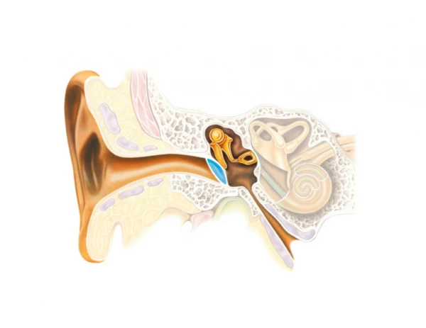
The middle ear Important Structures: • Epitympanic recess • Tympanic cavity • Aditus ad antrum • Mastoid air cells • Ossicles
The middle ear Schematic view of the middle ear boundaries and landmarks
The superior face • Tegmen Tympani
The inferior face • Tympanic plate • Jugular fossa
The medial face • Oval window • Footplate of the stapes • Round window • Promontory
The lateral face • Eardrum
The posterior face • Mastoid wall • Tympanic aditus • Pyramidal eminence • Chorda tympani nerve
The anterior face • Carotid wall • Eustachian tube
General structures of the middle ear • Eardrum • Ossicular chain • Eustachian tube • Middle ear muscles
The ossicles • Malleus (hammer) • Incus (anvil) • Stapes (stirrup)
The malleus • Manubrium • Neck • Head • Lateral process
The incus • Short process • Long process • Lenticular process • Incudostapedial joint
The stapes • Head • Neck • Anterior crus • Posterior crus • Footplate
Ligaments of the ossicular chain • Superior malleal ligament • Anterior malleal ligament • Lateral malleal ligament • Posterior incudal ligament
Purpose of the ossicuar chain • Impedance matching • Protection
Impedance matching of the middle ear • a sound wave traveling in a medium of certain physical properties, namely density and elasticity, will not pass readily into a medium with different properties • the more different the characteristics of the two media are, the more sound energy will be reflected at the boundary
Impedance matching of the middle ear • Acoustic resistance of air: 41.5 ohms • Acoustic resistance of cochlear fluid: 161,000 ohms • This represents a ratio of 3880:1 • Without the impedance matching capabilities of the middle ear, only 1/10 of 1% of the energy of an incoming sound wave would make it into the cochlea--99.9% of the energy would be reflected at the boundary
Area advantage • The area of the tympanic membrane is 17x the oval window • As the area decreases, the pressure increases
Impedance matching of the middle ear • Area advantage • Curved membrane buckling
Curved membrane buckling Notice how the eardrum curves from its rim at both ends to its attachment with the malleus in the middle. This point of the eardrum (V1) doesn’t move as far. This causes an increase in force.
Impedance matching of the middle ear • Area advantage • Curved membrane buckling • Lever action
Lever action advantage • The advantage is increased in (B) when the fulcrum is moved closer to the mass to be lifted.
Purpose of the ossicuar chain • Impedance matching • Protection
Purpose of the ossicuar chain The acoustic reflex Tensor tympani muscle Stapedius muscle
The tensor tympani • Larger of the two tympanic muscles • Tendon leaves the bony wall via the cochleariform process
The stapedius • The smaller of the two tympanic muscles • Tendon leaves the bony wall via the apex of the pyramidal eminence
The acoustic reflex • It is a reflex • Bilateral • Occurs in response to sound intensities delivered to either ear at 80-90 dB above threshold
The eustachian tube • 35-38 mm long • Oriented downward, forward, medialward • Osseous portion • Cartilaginous portion • Isthmus • Tensor palatini muscle