Download
1 / 65
680 likes | 951 Vues
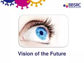
Vision of the Future. Keywords courtesy of www.wordle.net/. Develop an awareness of the importance of vision in our lives and the work that scientists do. Learning outcome. Vitamin D and healthy eyes.

E N D
Develop an awareness of the importance of vision in our lives and the work that scientists do. Learning outcome
Vitamin D and healthy eyes Vitamin D reduces the effects of ageing in mouse eyes and improves the vision of older mice. In the back of the eyes of mammals is a layer of tissue called the retina. Cells in the retina detect light as it comes into the eyes and then send messages to the brain, which is how we see. The retina has to have a good supply of blood. As we age we get more inflammation in the retina. Vitamin D reduces the inflammation.
Light, Body Clocks and Sleep An entirely new class of photoreceptor has been discovered - specialised cells found in the retina of the eye. The rods and cones detect light and transmit the information to our brains so we can use it to generate the image of the world we see. The new photoreceptors, called retinal ganglion cells, are specialised neurons that detect brightness. They regulate the timing of our internal 24-hour body clocks, sleep patterns, alertness, mood, blood pressure and even pupil size.
Vision and sport Does having excellent vision go hand in hand with elite sporting ability? Researchers have teamed up with the England & Wales Cricket Board to see if good vision is required for good hand-eye coordination. But do elite cricketers have superior vision to non-elites or novices? And if so, does this makes them good athletes or does their vision improve with training? The team will make detailed measures of visual function and relate these to performance on a specific aspect of cricket (one-handed catching).
Reindeer see a weird and wonderful world of ultraviolet light Researchers have discovered that reindeers have special vision that enables them to see ultraviolet (UV) light without it damaging their eyes. UV light causes snow blindness in humans. This ability is part of the reindeer's unique adaptation to the extreme arctic environment where they live. It allows them to see things vital for their survival.
Flies have the fastest vision Fly eyes have the fastest visual responses in the animal kingdom, but how they achieve this has long been an enigma. A fly's vision is so fast that it is capable of tracking movements up to five times faster than our own eyes. Researchers have discovered that this may be due to their photoreceptors physically contracting in response to light.
High-definition polarization vision discovered in cuttlefish Squid, octopus and cuttlefish are colour blind but can see aspects of light - including polarized light - that are invisible to humans. This gives them improved vision underwater and enables them to communicate in secret without other organisms being able to see their messages. Researchersdiscovered they have special high-definition vision by showing cuttlefish movies on modified LCD computer screens to test their eyesight. Researchers played the cuttlefish videos and watched for changes in skin colour patterns to determine if the cuttlefish could see small changes in the light.
Amazing cuttlefish facts Cuttlefish change the shape of the whole eye to move the position of the lens in order to focus; unlike humans who use our muscles to change the shape of the lens. They also have W shaped pupils and no blind spot, because the optic nerve is behind the retina.
The evolution of colour vision Being colour blind is often thought to be a disadvantage, but researchers have found that colour blind primates in the wild, as well as captivity, are better at catching camouflaged insects. Primates have three types of colour receptor in the retina known as cone cells. The receptors are often termed red, green and blue, which correspond to absorbance of light with long, medium and short wavelengths. Photopigments (opsins) produced in the cone photoreceptor cells absorb the light and trigger a signal that is sent to the brain.
Where is that nightjar? Animals use colours and patterns to blend into the background so they can remain hidden, but predators can learn to read this camouflage. Scientists are studying the different types of camouflage that exist in nature and how they hide prey animals from different types of predator vision. Scientists are using motion detecting cameras to study ground nesting birds in Zambia and South Africa, including nightjars. These birds, chicks and eggs have incredible camouflage but the scientists have discovered a variety of predators including vervet monkeys, mongooses and birds. To test their theories the scientists have developed computer games to find out how camouflage works. Can you find the nightjar? • You can play one of the games as either a mongoose or a monkey at http://nightjar.exeter.ac.uk/where-is-that-nightjar/
Reindeer see a weird and wonderful world of ultraviolet light Researchers have discovered that reindeers have special vision that enables them to see ultraviolet (UV) light without it damaging their eyes. UV light causes snow blindness in humans. This ability is part of the reindeer's unique adaptation to the extreme arctic environment where they live. It allows them to see things vital for their survival.
Learning outcomes Students will be able to: Identify internal and external anatomy of the eye, naming and locating the following parts: cornea, iris, pupil, lens, retina, sclera, choroid, optic nerve and blind spot. Relate the structure and function of the eye tissues . Investigate the properties of the eye tissues and their absorbance of UV light. Discuss eye diseases and disorders, such as snow blindness, describe how they occur, and name risk factors and possible preventative measures.
Checklist • Eye • Dissecting tray or cutting board • Forceps • Blunt probe • Sharp probe • Sharp pointed scissors (scalpels or razor) • Newspaper or paper towels • Eye protection • Apron • Rubbish bag • UV torch • UV exposure card
Stage 1 – Identification of external eye structure • Examine the outside of the eye. Locate the cornea at the front of the eye and the white sclera which is a tough outer layer surrounding the eye. The eye-lid may also still be attached. • The covering over the front of the eye is the cornea. The cornea is normally clear but turns cloudy after death. You may be able to see the iris, the coloured part of the eye, and the pupil, the dark oval in the middle of the iris behind the cornea.
Stage 1 – Identification of external eye structure • Surrounding the eye, particularly at the back, there should be fatty tissue which cushions the eye from shocks, an optic nerve and muscles that move the eyeball (indicated in images). These external muscles are termed extrinsic muscles, of which there are 4 in humans and 6 in sheep. You may need to move the muscles and fatty tissue aside to locate the optic nerve. • Carefully remove the eye-lid, fatty tissue and muscles from around the sclera and cornea taking care not to remove the optic nerve. • Label the parts you have identified on a diagram of the eye.
Stage 2 – Examining the internal anatomy of the eye • Place the eye down on the dissecting tray and turn it so the cornea faces to the left. You will need to make a vertical incision in the sclera half way between the cornea and the optic nerve to separate the front and rear of the eye. • Use the point of the scissors to make an initial cut. The scissors will need to be very sharp to cut through the tough outer covering. Care is required at this point as the eye may be very slippy and can be hard to hold in place while making the cut. • Once the cut is deep enough some fluid, the vitreous humour, may ooze out. The vitreous humour helps the eye maintain its shape and as it leaks out the eye may be harder to hold. • Insert the point of the scissors in the slit and cut around the eye with small snipping motions.
Stage 2 – Examining the internal anatomy of the eye • Rotate the eye as you cut until it has separated into the front (cornea) and rear (optic nerve) halves. Some of the vitreous humour is likely to leak out as you carry out this step. • Turn your attention to the front half of the eye. Using the forceps and blunt probe remove the rest of the vitreous humour taking care not to disturb the lens, ciliary body and suspensory ligaments. This may take some effort and require you to scrape the gooey fluid out. Try to save some on one side of your dissecting tray for later investigation. • Looking into the inside of the eye the lens will be visible as an oval structure held over the pupil by the suspensory ligaments. Remove the lens and note its shape, stiffness and degree of transparency. Some of the suspensory ligaments may remain attached. • Put the lens to one side for later investigations.
Stage 2 – Examining the internal anatomy of the eye • The pupil and iris should now be clearly visible. The size of the pupil (the opening that allows light to enter the eye) is controlled by two muscle layers in the iris. One layer increases the pupil size and the other layer reduces pupil size. In humans the pupil is circular but varies in shape in other organisms. • Between the iris and cornea is another cavity filled with a second semi-liquid fluid, the aqueous humour. This fluid, like the vitreous humour helps to maintain the shape of the eye. Use the point of the scissors to puncture a small slit at the boundary between the cornea and sclera. Then insert the scissors into the slip and cut all the way around the cornea. Remove the cornea from the front eye hemisphere. Notice the thickness of the cornea. • Put the cornea to one side for later investigation. • The iris should now be clearly visible from the front of the eye. Note the size of the pupil.
Stage 2 – Examining the internal anatomy of the eye • Now turn your attention to the rear half of the eye. Remove any remaining vitreous humour. On the inside of the back half of the eyeball, you can see some blood vessels that are part of a thin fleshy film. That film is the retina. Before you cut the eye open, the vitreous humour pushed against the retina so that it lay flat on the back of the eye. • Use your blunt probe to carefully lift and pull the retina back from the underlying choroid layer. Notice that the retina is only firmly attached to the choroid at one place. This region is the optic disc or blind spot. Here the nerve fibres from the photosensitive cells in the retina come together and form the optic nerve which carries signals to the brain. Recall identifying the optic nerve on the exterior of the eye at the start of the dissection. • Remove the retina and put it to one side for later investigation.
Stage 2 – Examining the internal anatomy of the eye • On the inside of the eye quite firmly attached to the sclera is a thin layer known as the choroid. Identify this layer and observe both its dark pigmentation and any shiny reflective properties. • Using your sharp probe insert the point between the choroid and sclera and moving around the back of the eye carefully remove the choroid from the sclera and place to one side for later investigation.
Stage 3 – Properties of eye tissues and UV absorbance • Place the lens on the UV card over some lettering. What do you observe? Note the change in the apparent size of the letters. This magnification is due to refraction. • Position the UV torch a short fixed distance away. A clamp stand can hold it in place. • Establish the minimum time required for maximum exposure that can be measured with the UV card using the UV light you have. • Place the dissected tissues of the eye one at a time over the circle on the UV card. • Shine the torch for the established length of time at the target area on the UV card.
Stage 3 – Properties of eye tissues and UV absorbance • Remove the tissue and record the amount of UV light that penetrated the tissue and was detected by the card. The results can be recorded with a camera or by taking quantitative measurements. • Repeat for the cornea, lens, vitreous humour, retina and choroid and establish the relative absorbance of UV light by the different tissues. • If you are comparing tissues from different organisms take into account the thickness of the tissues such as the lens. • Now that the tissues in the eye that absorb UV light have been investigated the effectiveness of sunglasses at protecting eyes from UV damage can be measured.
pupil – opening that lets light enter the eye cornea – bends light towards the pupil aqueous humour – transparent liquid behind the cornea iris – circular muscle that controls the size of the pupil, also contains coloured pigment Structure – Function
lens – adjusts its thickness to focus light on the retina ciliary muscle – changes shape of the lens suspensory ligaments – attaches ciliary muscle to eye covering Structure – Function
retina – inner layer of the eye that contains photoreceptors which detect light choroid layer – middle layer of the eye that contains blood vessels and nutrients for the eye sclera – white outer layer of the eye which protects it Structure – Function
vitreous humour – transparent jelly-like substance which fills the eyeball in to give its shape fovea centralis – very sensitive area of the retina for seeing fine detail optic nerve – nerve that carries impulses from eye to brain blind spot – area where optic nerve enters the eye, cannot see light focused here Structure – Function
Learning outcomes Students will be able to: Identify internal and external anatomy of the eye, naming and locating the following parts: cornea, iris, pupil, lens, retina, sclera, choroid, optic nerve and blind spot. Relate the structure and function of the eye tissues . Investigate the properties of the eye tissues and their absorbance of UV light. Discuss eye diseases and disorders, such as snow blindness, describe how they occur, and name risk factors and possible preventative measures.
Learning outcomes Students will be able to: Suggest the purpose of binocular vision Investigate hand eye coordination in relation to binocular vision Investigate the effect of decreased light level on the ability to perceive the distance of objects Discuss the effect of visual ability on sporting ability
Investigating binocular vision • Hold a pen or pencil at arms length in one hand. • Use one finger of the other hand to quickly touch the end of the pen. • Repeat this ten times using rapid movements to touch the pen. • Record the number of clean hits. • Repeat with one eye closed. • Hold two pens at arms length, one in each hand and with their points towards each other. • Touch the points together. • Now close one eye and try again. What difference does it make to use two eyes?
Illusory pendulum • Work in pairs • What you need: String, Plasticene, paper, pen, ruler, dark filter or a pair of sunglasses with one of the lenses removed. • Make a pendulum from a piece of string and ball of Plasticene. • Draw a straight line on a piece of paper using a ruler. • Practise swinging the pendulum in a straight arc across the line of sight in a regular rhythm. • Your partner should sit about two metres from the pendulum and observe the path of the pendulum as it swings from left to right and back again. • Your partner should then cover one eye with the dark filter. • Mark on the paper where the pendulum is swinging. • Your partner should record their observations and then swap over.
Testing binocular vision • Work in pairs • What you need: a tennis ball. • Stand facing your partner and determine a distance that you are reasonably confident you can reliably catch a tennis ball with one hand. Practice catching the ball and decide on a suitable distance. • Measure or mark out this distance to ensure you maintain this distance between throws. • The ball should be thrown ten times to your partner, recording the number of successful catches. After ten throws swap with your partner and repeat. • Repeat the activity with one eye covered, once again recording the number of successful catches. • Repeat the activity with the other eye covered. • Repeat the activity with a dark filter in front of one eye. Do you observe one eye to be better at detecting distance than the other?
Learning outcomes Students will be able to: Suggest the purpose of binocular vision Investigate hand eye coordination in relation to binocular vision Investigate the effect of decreased light level on the ability to perceive the distance of objects Discuss the effect of visual ability on sporting ability
Learning outcomes Students will be able to: identify their own level of colour discrimination investigate colour vision explain reflection, absorbance and filtering of light and the effect on colour