As Clinical Anatomy
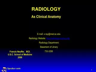
DIAGNOSTIC RADIOLOGY. Francis Neuffer, M.D. U.S.C. School of Medicine. As Clinical Anatomy. E-mail: x-ray@med.sc.edu Radiology Website: http://radiology.med.sc.edu Radiology Department: Basement of Library Penny Al-Emam- -216-3701. Speaker note.

As Clinical Anatomy
E N D
Presentation Transcript
DIAGNOSTIC RADIOLOGY Francis Neuffer, M.D. U.S.C. School of Medicine As Clinical Anatomy E-mail: x-ray@med.sc.edu Radiology Website: http://radiology.med.sc.edu Radiology Department: Basement of Library Penny Al-Emam--216-3701 Speaker note
COURSE GOALS • Understand basics of image generation. • Relate imaging to gross anatomy. • Appreciate indications and limitations. • Develop imaging vocabulary
WHAT IS DIAGNOSTIC RADIOLOGY? • 1 year internship • 4 year residency • 1 year fellowship?
RADIOLOGIST ROLE • Separate: Normal from Abnormal • Characterize / Describe: Abnormality • Determine: Extent (stage) of disease • Suggest: Diagnosis / Differential • Recommend: Further exams / follow-up
X-RAY • Discovered and named by Dr. W. C. Röentgen at University of Würzburg, 1895 • Awarded first Nobel prize for physics, 1901 • Did not patent invention
FOOT AP PROJECTION (ANTERIOR – POSTERIOR) * * =
TOMOGRAPHIC IMAGES ARE IN A SPECIFIC PLANE AXIAL CORONAL SAGITTAL RT RT
CT- HEAD RT CT REFERENCE FILM Skull / brain
RADIOLOGY TOOLS X- RAY ULTRASOUND NUCLEAR MEDICINE MAGNETIC RESONANCE COMPUTED TOMOGRAPHY
HOW IS IMAGING DONE? • IONIZING RADIATION X-ray, CT, Nuclear Medicine • SOUND WAVES Ultrasound • MAGNETIC FIELDS / RADIO WAVES Magnetic Resonance
X-RAY • High Energy Photon --Kilo Electron Volts • Ionizing Radiation • Exposes Film / Detector • Projection Data X-ray beam detector
X-RAYSPLAIN FILM RADIOGRAPHY • Chest • Mammography • Abdomen • Spine • Extremities & Joints • Skull
X - RAY --- FOUR BASIC DENSITIES • Air • Soft Tissue • Bone • Fat
CONTRAST RADIOGRAPHY • Injection, ingestion, or other placement of opaque material within the body. • Improves visualization and tissue separation. • Can demonstrate functional anatomy and pathology.
UPPER GI--(GASTRO INTESTINAL) STOMACH ORAL BARIUM CONTRAST WITHOUT CONTRAST-plain or scout film COLON BARIUM ENEMA RECTAL BARIUM CONTRAST
INTRAVENOUS PYELOGRAM – IVP INTRAVENOUS IODINE CONTRAST WITHOUT CONTRAST-plain or scout film ARTERIOGRAM INTRAARTERIAL IODINE CONTRAST
COMPUTED TOMOGRAPHY • HIGH ENERGY PHOTON • IONIZING RADIATION • EXPOSES DETECTOR • TOMOGRAPHIC DATA
CT EXAMPLE SCAN LEVEL Air Fat Soft tissue RT Bone
NUCLEAR MEDICINE • High Energy Photon • Ionizing Radiation --Radiopharmaceutical • Exposes Detector • Projection Data • Dynamic / Physiologic
Bone NUCLEAR MEDICINE EXAMPLES • Liver • PET scan
ULTRASOUND • Sound Wave - high Frequency-megahertz • No Ionizing Radiation • Tomographic Data
BASIC ULTRASOUND PHYSICSUltrasound Production • “Piezoelectric Effect” • Transducer/Probe 25
BASIC ULTRASOUND PHYSICSUltrasound Production • Transducer is the “speaker” &“microphone” • 99% of time is spent “listening” • Only 1% of time is devoted to making ultrasound 26
BASIC ULTRASOUND PHYSICSAcoustic Windows • Dense & elastic structures • Liver • Spleen • Fluid-filled structures • Heart • Urinary bladder 27
B Mode-brightness • Most common use • Presents “real time” image • Ultrasound Sector Scanning 28
ULTRASOUNDideal for fluid filled structures Gallbladder Kidney Obstetrics
MAGNETIC RESONANCE • Hydrogen Protons In a Magnetic Field • Radio Wave Signal Transmission • No Ionizing Radiation • Tomographic Data
MAGNETIC RESONANCEEXAMPLES Anterior RT • Brain • Spine Posterior Anterior Posterior
RADIOLOGY EVALUATION • Multiple Choice - Identify • Labeled Images From Digital Film Sets And Lecture Images
HOSPITAL LINGO You will hear and see these abbreviations used frequently in the medical community. X- Ray Plain Film Scout Film Radiograph Computed Tomography Cat Scan CT Nuclear Medicine Nuc Med Ultrasound Sono Sonogram Magnetic Resonance MR MRI
SUMMARY • TOMOGRAPHY- VS- PROJECTION IMAGES • SECTION PLANES AXIAL CORONAL SAGITTAL • IMAGE GENERATION OF: Nuclear Medicine Computed Tomography Ultrasound X-ray Magnetic Resonance
OH… THE MANY USES OF NUC MED STUDY OF A HORSE’S _____! KING TUT MODERN IMAGING?