Lens diseases
Lens diseases. Wen Xu Eye Center, 2nd Affiliated Hospital Zhejiang University. The most common cause of painless, progressive loss of vision today is. ?. Cataract. Anatomy of the lens. Location posterior to iris anterior to vitreous Shape biconvex Structure lens capsule

Lens diseases
E N D
Presentation Transcript
Lens diseases Wen Xu Eye Center, 2nd Affiliated Hospital Zhejiang University
The most common cause of painless, progressive loss of vision today is ?
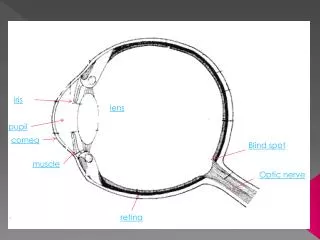
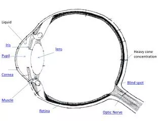
Anatomyofthelens • Location posterior to iris anterior to vitreous • Shape biconvex • Structure lens capsule lens cortex lens nucleus
Physiology of the lens • No vessel, nerve and transparent. • Derive nutrients from the aqueous humor • Significant refractive medium • Accommodative function • No immediate relation with adjacent tissues • Complex metabolism • Simple disorders: transparency and location change
Cataract • Definition: opacification of the lens • epidemiology: clinical cataract corrected vision<0.5
Cataract Mechanism: many factors lens capsular damage osmosis increase,loss of protective screen,metabolic disorders protein degeneration,cell apoptosis lens opacify cataract
Cataract Classification: • by cause: congenital, senile(age-related), complicated, metabolitic, drug-induced, toxic, traumatic, secondary • by age:congenital, acquired • by location: cortical, nuclear, subcapsular • by shape: dot-like, coronary, lamellar • by degree: immature, intumescent, mature, hypermature
Cataract Symptoms: • decreased vision: most obvious and important • decreased contrast sensitity • refractive error: myopia,astigmatism • monocular diplopia or multiple vision • glare: scattered light rays • poor color discrimination:blue spectrum
Cataract Signs: The lens is best examined with the pupil dilated. A magnified view of the lens can be obtained with a slit-lamp or by using the direct ophthalmoscope with a high plus (10+) setting
Cataract Grade’s standards of nuclear hardness: Ⅰ transparent,no nucleus,soft Ⅱ yellow-white or yellow,soft Ⅲ dark yellow,moderate hard Ⅳ brown or amber, hard Ⅴ brown or black,extremely hard
(I) Age-related cataract Description: the most common type, most patients are beyond their 50’s. The incidence goes up with aging. It is the first rank of ophthalmic diseases leading to blindness Risk factors: Many factors are involved include age, occupation, sex, ultraviolet radiation, diabetes, hypertension, positive family history, nutritious condition
(i) Cortical Cataract • The most common type • Four stages: (1) incipient stage (2) intumescent stage or immature stage (3) mature stage (4) hypermature stage
1. incipient stage Features: a、cuneiform(楔形['kjunɪə,fɔrm]) opacity b、lamellar seperate c、vacuole d、cracks e、no vision damage Tests:a、slit-lamp b、transillumination
2. intumescent stage or immature stage Features: a、more serious opacity b、larger volume and more shallow anterior chamber c、iris shadow d、obvious vision decrease e、myopia Tests:a、slit-lamp b、oblique illumination Matter needs attention: angle-closure glaucoma
3. mature stage Features: a、complete opacity, milky white, iris shadow disappear b、volumn and anterior chamber regain normal c、vision: LP or HM before the eye Tests:a、slit-lamp b、flashlight
4. hypermature stage Features: a、smaller volumn,wrinkled lens capsule,claybank and fallen nucleus (Morgagnian cataract) ,superior of anterior chamber deepens while inferior is the opposite,ridodonesis. b、laceration of lens capsule,lens luxation. c、phacoanaphylactic uveitis,phacolytic glaucoma
(ii) Nuclear Cataract Features: a、start earlier,generally on 40’s,slowly progressive, not likely to be mature. b、nuclear opacity: start by embryonic nucleus. c、vision: no vision damage early on, myopia Tests:slit-lamp、 transillumination、 oblique illumination
(iii) Subcapsular Cataract Features: a、start earlier b、posterior subcapsular cataract: cause obvious vision defect early on c、cupuliform(杯状) opacity of posterior pole
(II) Congenital Cataract Features: present at birth or appear shortly thereafter; unilateral or bilateral; may be alone or associated with other ocular or systemic congenital abnomalities Etiology: (1) hereditary factors(chromosome,gene) (2) environmental factors (matrix disease) when pregnance <3 m: virus infection; drugs,metabolic diseases (3) undetermined causes
Classification According to location, form and degree • anterior polar cataract • posterior polar cataract • perinuclear cataract • coronary cataract • punctate cataract • total cataract • membrane cataract • nuclear cataract
(III) complicated cataract Features: ocular inflammation or degenerative disorders→ nutritious or metabolic defect → lens opacity Common causes: corneal ulcer, glaucoma, uveitis,retinal detachment, retinitis pigmentosa, intraocular tumor,high myopia, etc.
Clinical findings: 1. primary disease changes 2. cataract Treatment: 1. treat the primary disease. 2.do the surgery after 3 m of inflammation control
(IV) Metabolic cataract • Diabetic cataract • Galactose cataract: lack of enzyme • Tetany cataract: low blood calcium • Wilson’s disease (Hepatolenticular Degeneration): KF ring, sunflower-shaped opacity,copper.
1. Diabetic cataract • Mechanism: blood sugar↑ →sugar in the lens↑ → change into sorbitol→plasma osmotic pressure↑ →absorb water→fibers swellen and degenerate→lens opacity
classification: (1) real diabetic cataract (2) age-related cataract of diabetic patients • Clinical findings: (1) the first type: teenagers,bilateral,rapidly progressive,eading to total cataract,combined with refractive changes according to blood sugar (2) the second type: high incidence,start earlier, fast progressive, easy to be mature,similar with senile cortical cataract
Treatment: (1) positively treat diabetes,control blood sugar (2) do the surgery if permitted (3) positively postoperational infection and bleeding prevention
(V) Drug-induced and toxic cataract • Corticosteroid cataract • Chlorpromazine cataract • Miotic cataract • Trinitrotoluence cataract • Metals
(VI) Traumatic cataract • Classification: Contusive cataract Penetrating cataract Chemical injuries cataract Radiation cataract Electric cataract • Treatment: observation or surgery
(VII) Secondary cataract • Definitions: opacification of the posterior capsule due to partially absorbed traumatic cataract or following extracapsular cataract extraction (posterior capsular opacification). It is the most common complication of cataract surgery • Clinical findings: vision decrease after cataract surgery; Elschnig’s pearls
It is a significant problem in almost all pediatric patients unless the posterior capsule and anterior vitreous are removed at the time of surgery. Up to 30%-50% of all adult patients develop an opaque secondary membrane after cataract surgery • Treatment: neodymium:YAG capsulotomy