Download
1 / 98
1.02k likes | 2.5k Vues
s and Femoral Head Fractures. INJURIES AROUND HIP {Hip dislocations and Head fractures}. Introduction. Hip dislocations caused by significant force: Association with other fractures Damage to vascular supply to femoral head Thus, high chance of complications. Anatomy: Hip Joint.

E N D
s and Femoral Head Fractures • INJURIES AROUND HIP • {Hip dislocations and Head fractures}
Introduction Hip dislocations caused by significant force: • Association with other fractures • Damage to vascular supply to femoral head Thus, high chance of complications
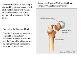
Anatomy:Hip Joint Ball and socket joint. Femoral head: slightly asymmetric, forms 2/3 sphere. Acetabulum: inverted “U” shaped articular surface. Ligamentum teres, with artery to femoral head, passes through middle of inverted “U”.
Joint Contact Area Throughout ROM: • 40% of femoral head is in contact with acetabular articular cartilage. • 10% of femoral head is in contact with labrum.
Acetabular Labrum Strong fibrous ring Increases femoral head coverage Contributes to hip joint stability
Hip Joint Capsule • Extends from intertrochanteric ridge of proximal femur to bony perimeter of acetabulum • Has several thick bands of fibrous tissue Iliofemoral ligament • Upside-down “Y” • Blocks hip hyperextension • Allows muscle relaxation with standing
Femoral Neck Anteversion • Averages 70 in Caucasian males. • Slightly higher in females. • Asian males and females have been noted to have anteversion of 140 and 160 respectively.
Blood Supply to Femoral Head • Artery of Ligamentum Teres • Most important in children. • Its contribution decreases with age, and is probably insignificant in elderly patients.
Blood Supply to Femoral Head 2. Ascending Cervical Branches • Arise from ring at base of neck. • Ring is formed by branches of medial and lateral circumflex femoral arteries. • Penetrate capsule near its femoral attachment and ascend along neck. • Perforate bone just distal to articular cartilage. • Highly susceptible to injury with hip dislocation.
Sciatic Nerve Composed from roots of L4 to S3. Peroneal and tibial components differentiate early, sometimes as proximal as in pelvis. Passes posterior to posterior wall of acetabulum. Generally passes inferior to piriformis muscle, but occasionally the piriformis will split the peroneal and tibial components
Posterior Hip Dislocation: Mechanism of Injury Almost always due to high-energy trauma. Most commonly involve unrestrained occupants in MVAs. Can also occur in pedestrian-MVAs, falls from heights, industrial accidents and sporting injuries.
Posterior Dislocation • Generally results from axial load applied to femur, while hip is flexed. • Most commonly caused by impact of dashboard on knee.
Type of Posterior Dislocation depends on: Direction of applied force. Position of hip. Strength of patient’s bone.
Hip Position vs. Type of Posterior Dislocation In General, Abduction: acetabulum fracture-dislocation Adduction: pure dislocation Extension: femoral head fracture-dislocation Flexion: pure dislocation
Anterior Dislocation 7-10% of hip dislocations Mechanism: • Forced abduction with external rotation of hip. • Anterior hip capsule is torn or avulsed. • Femoral head is levered out anteriorly.
Effect of Dislocation on Femoral Head Circulation • Injury to ascending cervical branches associated with damage to capsule during dislocation. • Dislocation disrupts artery of ligamentum teres. • Dislocated hip may kink or compress acending cervical branches until the hip is reduced. Thus, early reduction of the dislocated hip can improve blood flow to femoral head.
Associated Injuries Mechanism: knee vs. dashboard Contusions of distal femur Patella fractures Foot fractures, if knee extended
Associated Injuries Sciatic nerve injuries occur in 10% of adult and 5% of pediatric hip dislocations. Most commonly, these resolve with reduction of hip and passage of time. Stretching or contusion most common. Piercing or transection of nerve by bone can occur.
Thompson and Epstein Classificationof Hip Dislocations (Most well-known) Type I Pure dislocation with at most a small posterior wall fragment. Type II Dislocation with large posterior wall fragment. Type III Dislocation with comminuted posterior wall. Type IV Dislocation with “acetabular floor” fracture (probably transverse + post. wall acetabulum fracture-dislocation). Type V Dislocation with femoral head fracture. Thompson and Epstein, J Bone and Joint Surg, 1951
Epstein Classificationof Anterior Hip Dislocations Type I Superior (pubic and subspinous) Type II Inferior (obturator and perineal) A No associated fracture B Associated fracture of the femoral head/neck C Associated fracture of the acetabulum i.e, Type IA, IIB, etc. Epstein, Clin Orthop Relat Res, 1973.
AO/OTA Classification • Most thorough. • Best for reporting data, to allow comparison of patients from different studies. • 30-D10 Anterior Hip Dislocation • 30-D11 Posterior Hip Dislocation • 30-D30 Obturator (Anterior-Inferior) Hip Dislocation
Evaluation: History Significant trauma, usually MVA. Awake, alert patients have severe pain in hip region.
Physical Examination: Classical Appearance Posterior Dislocation: Hip flexed, internally rotated, adducted.
Physical Examination: Classical Appearance Anterior Dislocation: Extreme external rotation, less-pronounced abduction and flexion.
Unclassical presentation (posture) if: • femoral head or neck fracture • femoral shaft fracture • obtunded patient
Physical Examination • Pain to palpation of hip. • Pain with attempted motion of hip. • Possible neurological impairment: Thorough exam essential!
Radiographs: AP Pelvis X-Ray • In primary survey of ATLS Protocol. • Should allow diagnosis and show direction of dislocation. • Femoral head not centered in acetabulum. • Femoral head appears larger (anterior) or smaller (posterior). • Usually provides enough information to proceed with closed reduction.
Reasons to Obtain More X-Rays Before Hip Reduction • View of femoral neck inadequate to rule out fracture. • Patient requires CT scan of abdomen/pelvis for hemodynamic instability • and additional time to obtain 2-3 mm cuts through acetabulum + femoral head/neck would be minimal.
X-rays after Hip Reduction: • AP pelvis, Lateral Hip x-ray. • Judet views of pelvis. • CT scan with 2-3 mm cuts.
CT Scan Most helpful after hip reduction. Reveals: Non-displaced fractures. Congruity of reduction. Intra-articular fragments. Size of bony fragments.
MRI Scan • Will reveal labral tear and soft-tissue anatomy. • Has not been shown to be of benefit in acute evaluation and treatment of hip dislocations.
Clinical Management: Emergent Treatment • Dislocated hip is an emergency. • Goal is to reduce risk of AVN and DJD. • Evaluation and treatment must be streamlined.
Emergent Reduction • Allows restoration of flow through occluded or compressed vessels. • Requires proper anesthesia. • Requires “team” (i.e. more than one person).
Anesthesia • General anesthesia with muscle relaxation facilitates reduction, but is not necessary. • Conscious sedation is acceptable. • Attempts at reduction with inadequate analgesia/ sedation will cause unnecessary pain, create muscle spasm, and make subsequent attempts at reduction more difficult.
Reduction Maneuvers Allis: Patient supine. Requires at least two people. Stimson: Patient prone, hip flexed and leg off stretcher. Requires one person. Impractical in trauma (i.e. most patients).
Allis Maneuver • Assistant: Stabilizes pelvis • Posterior-directed force on both ASIS’s • Surgeon: Stands on stretcher • Gently flexes hip to 900 • Applies progressively increasing traction to the extremity • Applies adduction with internal rotation • Reduction can often be seen and felt
Reduced Hip • Moves more freely • Patient more comfortable • Requires testing of stability • Simply flexing hip to 900 does not sufficiently test stability
Stability Test • Hip flexed to 90o • If hip remains stable, apply internal rotation, adduction and posterior force. • The amount of flexion, adduction and internal rotation that is necessary to cause hip dislocation should be documented. • Caution!: Large posterior wall fractures may make appreciation of dislocation difficult.
Irreducible Hip Requires emergent reduction in O.R. Pre-op CT obtained if it will not cause delay. One more attempt at closed reduction in O.R. with anesthesia. • Repeated efforts not likely to be successful and may create harm to the neurovascular structures, articular cartilage, or even cause iatrogenic fracture. Stannard et al, Clin Orthop Relat Res, 2000 Surgical approach from side of dislocation.
Hip Dislocation: Nonoperative Treatment • If hip stable after reduction, and reduction congruent. • Maintain patient comfort. • ROM precautions (No Adduction, Internal Rotation). • No flexion > 60o. • Early mobilization. • Touch down weight-bearing for 4-6 weeks. • Repeat x-rays before allowing weight-bearing.
Hip Dislocation:Indications for Operative Treatment • Irreducible hip dislocation • Hip dislocation with femoral neck fracture • Incarcerated fragment in joint • Incongruent reduction • Unstable hip after reduction
1. Irreducible Hip Dislocation: Anterior Smith-Peterson approach • Watson-Jones is an alternate approach • Allows visualization and retraction of interposed tissue. • Placement of Schanz pin in intertrochanteric region of femur will assist in manipulation of the proximal femur. • Repair capsule, if this can be accomplished without further dissection.
1. Irreducible Hip Dislocation: Posterior • Kocher-Langenbeck approach. • Remove interposed tissue, or release buttonhole. • Repair posterior wall of acetabulum if fractured and amenable to fixation.
Irreducible Posterior Dislocation with Large Femoral Head Fracture Fortunately, these are rare. Difficult to fix femoral head fracture from posterior approach without transecting ligamentum teres.
Three Options • Detach femoral head from ligamentum teres, repair femoral head fracture with hip dislocated, reduce hip. • Reduce hip through posterior incision, close posterior wound, fix femoral head fracture from anterior approach (either now or later). • Ganz trochanteric “flip” osteotomy. Best option not known: Damage to blood supply from anterior capsulotomy vs. damage to blood supply from transecting ligamentum teres. These will be discussed in detail in femoral head fracture section.
2. Hip Dislocation with Femoral Neck Fracture Attempts at closed reduction potentiate chance of fracture displacement with consequent increased risk of AVN. If femoral neck fracture is already displaced, then the ability to reduce the head by closed means is markedly compromised. Thus, closed reduction should not be attempted.
2. Hip Dislocation with Femoral Neck Fracture Usually the dislocation is posterior. Thus, Kocher-Langenbeck approach. If fracture is non-displaced, stabilize fracture with parallel lag screws first. If fracture is displaced, open reduction of femoral head into acetabulum, reduction of femoral neck fracture, and stabilization of femoral neck fracture.
3. Incarcerated Fragment Can be detected on x-ray or CT scan. Surgical removal necessary to prevent abrasive wear of the articular cartilage. Posterior approach allows best visualization of acetabulum (with distraction or intra-op dislocation). Anterior approach only if: dislocation was anterior and, fragment is readily accessible anteriorly.
4. Incongruent Reduction Caused By: • Acetabulum Fracture (weight-bearing portion). • Femoral Head Fracture (any portion). • Interposed tissue. Goal: achieve congruence by removing interposed tissue and/or reducing and stabilizing fracture.