Download
1 / 44
1.04k likes | 3.04k Vues
Nuclear Magnetic Resonance (NMR) Spectroscopy. Prof. Yonghai Chai. School of Chemistry & Materials Science. For Bilingual Chemistry Education. Definition of NMR Spectroscopy.

E N D
Nuclear Magnetic Resonance (NMR) Spectroscopy Prof. Yonghai Chai School of Chemistry & Materials Science For Bilingual Chemistry Education
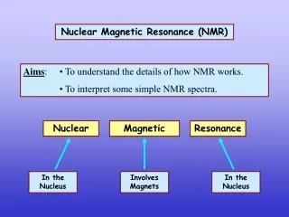
Definition of NMR Spectroscopy Nuclear magnetic resonance spectroscopy: commonly referred to as NMR,is a technique which exploits the magnetic properties of certain nuclei to study physical, chemical, and biological properties of matter Compared to mass spectrometry, larger amounts of sample are needed, but non-destructive
NMR History • 1937 Rabi’s prediction and observation of nuclear magnetic resonance • 1945 First NMR of solution (Bloch et al for H2O) and solids (Purcell et • al for parafin)! • 1953 Overhauser NOE (nuclear Overhauser effect) • 1966 Ernst, Anderson Fourier transform NMR • 1975 Jeener, Ernst 2D NMR • 1980 NMR protein structure by Wuthrich • 1990 3D and 1H/15N/13C Triple resonance • 1997 Ultra high field (~800 MHz) & TROSY(MW 100K)
Continuation of NMR History Nobel prizes 1944 Physics Rabi (Columbia) 1991 Chemistry Ernst (ETH) 1952 Physics Bloch (Stanford), Purcell (Harvard) "for his resonance method for recording the magnetic properties of atomic nuclei" "for their development of new methods for nuclear magnetic precision measurements and discoveries in connection therewith" "for his contributions to the development of the methodology of high resolution nuclear magnetic resonance (NMR) spectroscopy"
Continuation of NMR History 2002 Chemistry Wüthrich (ETH) "for his development of nuclear magnetic resonance spectroscopy for determining the three-dimensional structure of biological macromolecules in solution" 2003 Medicine Lauterbur (University of Illinois in Urbana ), Mansfield (University of Nottingham) "for their discoveries concerning magnetic resonance imaging"
Spin of Nuclei Fermions : Odd mass nuclei with an odd number of nucleons have fractional spins. I = 1/2 ( 1H, 13C, 19F, 31P ), I = 3/2 ( 11B, 33S ) & I = 5/2 ( 17O ). Bosons : Even mass nuclei with odd numbers of protons and neutrons have integral spins. I = 1 ( 2H, 14N ) Even mass nuclei composed of even numbers of protons and neutrons have zero spin I = 0 (12C, and 16O, 32S)
Angular Momentum A spinning charge generates a magnetic field, the resulting spin-magnet has a magnetic moment (μ) proportional to the spin I magnetic moment (磁矩) m = gp whereg is the gyromagnetic ratio (旋磁比), and it is a constant for a given nucleus When I=0, m=0 ** There is no spin for nuclei with I=0 • “Right Hand Rule” • determines the direction of the magnetic field around a current-carrying wire and vice-versa
Energy Differentiation In the presence of an external magnetic field (B0), two spin states exist, +1/2 and -1/2 (For I=1/2).The magnetic moment of the lower energy +1/2 state is aligned with the external field, and that of the higher energy -1/2 spin state is opposed to the external field. Aligned against the applied field Aligned with the applied field
Energy Differentiation • Difference in energy between the two states is given by: • DE = g h Bo / 2p • where: • Bo – external magnetic field • h – Planck’s constant • g–gyromagnetic ratio When the energy of the photon matches the energy difference between the two spin states , an absorption of energy occurs. We call that phenomenon Resonance DE = hu = ghBo / 2pSo,u = g Bo / 2p
Larmor Precession • Spinning particle precesses about the external field axis with an angular frequency known as the Larmor frequency wL = g Bo When radio frequency energy matching the Larmor frequency is introduced at a right angle to the external field, it would cause a transition between the two energy levels of the spin. In other world, the precessing nucleus will absorb energy and the magnetic moment will flip to its I = _1/2 state
Fourier transformation and the NMR spectrum Fourier transform RF Pulse The Fourier transform (FT) is a computational method for analyzing the frequencies present in an oscillating signal The NMR spectrum
1H NMR and 13C NMR Spectrum 1H NMR spectra d ppm High field Down field 13C NMR spectra d ppm
Chemical Shift-d When an atom is placed in a magnetic field, its electrons circulate about the direction of the applied magnetic field. This circulation causes a small magnetic field at the nucleus which opposes the externally applied field The magnetic field at the nucleus (the effective field) is therefore generally less than the applied field by a fraction : B = B0 (1-s), So u = g B0 (1-s) / 2p
Chemical Shift-d The electron density around each nucleus in a molecule varies according to the types of nuclei and bonds in the molecule. The opposing field and therefore the effective field at each nucleus will vary. This is called the chemical shift phenomenon. As we can tell from n = g B0 (1-s) / 2p, the greater the value of Bo, the greater the frequency difference. This relationship could make it difficult to compare NMR spectra taken on spectrometers operating at different field strengths. The term chemical shift was developed to avoid this problem. The chemical shift of a nucleus is the difference between the resonance frequency of the nucleus and a standard, relative to the standard. This quantity is reported in ppm and given the symbol delta. d = (n - nref) x106 / nref
Standard for Chemical Shift In NMR spectroscopy, the standard is often tetramethylsilane, Si(CH3)4, abbreviated TMS. Tetramethyl silane (TMS) is used as reference because it is soluble in most organic solvents, is inert, volatile, and has 12 equivalent 1H and 4 equivalent 13C. TMS signal is set to 0
Shielding and Deshielding A nucleus is said to be shieldedwhen electrons around the nucleus circulates in a magnetic field and create a secondary induced magnetic field which opposes the applied field . Trends in chemical shift are explained based on the degree of shielding or deshielding , e.g. of deshielding effect
Chemical Shift-d • Chemical shift depends on : • Electronegativity of nearby atoms • Hybridization of adjacent atoms • diamagnetic effects • paramagnetic effects • solvent effect
Spin-Spin Coupling Spin-spin coupling: The coupling of the intrinsic angular momentum of different particles. Such coupling between pairs of nuclear spins is an important feature of nuclear magnetic resonance (NMR) spectroscopy as it can provide detailed information about the structure and conformation of molecules. Spin-spin coupling between nuclear spin and electronic spin is responsible for hyperfine structure in atomic spectra.
J-Coupling J-coupling: also called indirect spin-spin coupling, is the coupling between two nuclear spins due to the influence of bonding electrons on the magnetic field running between the two nuclei. J-coupling provides information about dihedral angles, which can be estimated using the Karplus equation. It is an important observable effect in 1D NMR spectroscopy. The coupling constant, J (usually in frequency units, Hz) is a measure of the interaction between a pair of nuclei
1H-NMR • 1H experiencing the same chemical environment • or chemical shift are called equivalent hydrogens. • 1H experiencing different environment or having • different chemical shifts are nonequivalent hydrogens.
Factors to Affect 1H Chemical Shift Chemical shift : (1) electronegativity of nearby atoms, (2) hybridization of adjacent atoms, and (3) diamagnetic effects Electronegativity
Carbon-Carbon Triple Bond Effect A carbon-carbon triple bond shields an acetylenic hydrogen and shifts its signal to lower frequency (to the right) to a smaller value
Carbon-Carbon Double Bond Effect Magnetic induction in the p bond of a carbon-carbon double bond deshieldsvinylic hydrogens and shifts their signal higher frequency
Aromatic Effect The magnetic field induced by circulation of p electrons in an aromatic ring deshields the hydrogens on the ring and shifts their signal to higher frequency
Signal Splitting for 1H Peak: The units into which an NMR signal is split; doublet, triplet, quartet, multiplet, etc. Signal splitting: Splitting of an NMR signal into a set of peaks by the influence of neighboring nonequivalent hydrogens. (n + 1) rule: If a hydrogen has n hydrogens nonequivalent to it but equivalent among themselves on the same or adjacent atom(s), its 1H-NMR signal is split into (n + 1) peaks.
Pascal’s triangle The relative peak intensities for multiplet peaks arising from J- coupling of a 1H to N equivalent 1H can be determined using Pascal’s triangle:
Coupling constant Coupling constant (J): The separation on an NMR spectrum (in hertz) between adjacent peaks in a multiplet.
13C-NMR Spectroscopy Organic compounds contain carbon. Unfortunately, the C-12 nucleus does not have a nuclear spin, but the C-13 nucleus does due to the presence of an unpaired neucarbon-1tron. C-13 nuclei make up approximately 1% of the carbon nuclei on earth. Therefore, 13C NMR will be much less sensitive than 1HNMR NMR
13C-NMR Spectroscopy The presence of spin-spin coupling between a 13C nucleus and the nuclei of 1H atoms bonded to the 13C, splits the carbon-13 peaks and causes an even poorer signal-to-noise ratio • Each nonequivalent 13C gives a different signal • A 13C signal is split by the 1H bonded to it according to the (n + 1) rule. • Coupling constants of 100-250 Hz are common, which means that there is often significant overlap between signals, and splitting patterns can be very difficult to determine. • The most common mode of operation of a 13C-NMR spectrometer is a proton-decoupled mode.
Decoupling proton-decoupled mode, a sample is irradiated with two different radiofrequencies. One to excite all 13C nuclei, a second to cause all protons in the molecule to undergo rapid transitions between their nuclear spin states. On the time scale of a 13C-NMR spectrum, each proton is in an average or effectively constant nuclear spin state, with the result that 1H-13C spin-spin interactions are not observed and they are decoupled.
Chemical Shift - 13C-NMR • Trends • RCH3 < R2CH2 < R3CH • Electronegative atoms cause downfield shift • Pi bonds cause downfield shift • C=O 160-210 ppm
13C-NMR: Integration time for nucleus to relax from excited spin state to ground state • 1H-NMR: Integration reveals relative number of hydrogens per signal • 13C-NMR: Integration reveals relative number of carbons per signal • Rarely useful due to slow relaxation time for 13C
Interpreting NMR Spectra • Alkanes • 1H-NMR signals appear in the range of 0.8-1.7. • 13C-NMR signals appear in the considerably wider range of 10-60. • Alkenes • 1H-NMR signals appear in the range 4.6-5.7. • 1H-NMR coupling constants are generally larger for trans-vinylic hydrogens (J= 11-18 Hz) compared with cis-vinylic hydrogens (J= 5-10 Hz). • 13C-NMR signals for sp2 hybridized carbons appear in the range 100-160, which is to higher frequency from the signals of sp3 hybridized carbons.
Interpreting NMR Spectra Alcohols 1H-NMR O-H chemical shift often appears in the range 3.0-4.0, but may be as low as 0.5. 1H-NMR chemical shifts of hydrogens on the carbon bearing the -OH group are deshielded by the electron-withdrawing inductive effect of the oxygen and appear in the range 3.0-4.0. Ethers A distinctive feature in the 1H-NMR spectra of ethers is the chemical shift, 3.3-4.0, of hydrogens on the carbons bonded to the ether oxygen.
b b a a
Interpreting NMR Spectra • Aldehydes and ketones • 1H-NMR: aldehyde hydrogens appear at 9.5-10.1. • 1H-NMR: a-hydrogens of aldehydes and ketones appear at 2.2-2.6. • 13C-NMR: carbonyl carbons appear at 180-215. • Amines • 1H-NMR: amine hydrogens appear at 0.5-5.0 depending on conditions.
b a c c b c a b c a a 1H NMR isobutyraldehyde b 1H NMR Methyl ethyl ketone
Interpreting NMR Spectra • Carboxylic acids • 1H-NMR: carboxyl hydrogens appear at 10-13 ppm, higher than most other types of hydrogens. • 13C-NMR: carboxyl carbons in acids and esters appear at 160-180 ppm. b a c a c b