UROLOGICAL EMERGENCIES
UROLOGICAL EMERGENCIES. Julian Mander. Emergencies. Renal colic diagnosis and management Urine Retention Urosepsis Haematuria Testicular torsion Trauma renal bladder urethral. Stones - Presentation - Pain. PAIN

UROLOGICAL EMERGENCIES
E N D
Presentation Transcript
UROLOGICAL EMERGENCIES Julian Mander
Emergencies • Renal colic diagnosis and management • Urine Retention • Urosepsis • Haematuria • Testicular torsion • Trauma renal bladder urethral
Stones - Presentation - Pain PAIN Typical: Loin to groin pain Variable severity Episodic Not mechanical but paroxysmal Atypical: Anterior Groin pain alone Testicular pain alone Penile tip pain alone Associated vomiting (ongoing) Mechanical character
Stones - Presentation - LUTS LUTS Irritative: Frequency Urgency Strangury Burning micturition Macroscopic haematuria Common misdiagnosis UTI - Do MSUs if in doubt.
Stones - Presentation - Fever FEVER Loin pain + fever (38c) = pyelonephritis = generally not life threatening Loin pain + fever + stone = infected obstructed kidney = commonly life threatening Message – do ultrasound in all pyelonephritis admissions
Stones - Investigation MSU U&E/Creatinine Serum calcium/albumin uric acid Imaging First or infrequent presentation admit to hospital until diagnosis made
Diagnostic Imaging Until recently, limited IVP with extra tomograms or delayed films as dictated by progress Now imaging by non contrast stone CT scan as routine initial diagnostic imaging protocol. BUT CT scan has a relatively high radiation dose SO 1) Do not repeat CT for the same stone – once a diagnosis is established, patients can be managed with AXR or U/S or both. 2) Do not do CT scans for recurrent stone formers – patients can usually tell you the diagnosis – do U/S and AXR not CT. 3) Avoid CT in children 4) Do not do CT in pregnancy – use U/S
CT vs IVP Renal colic: A prospective evaluation of non-enhanced spiral CT versus intravenous pyelography Mendelson et al Australasian Radiology 2003 47, 22 – 28 200 patients randomized to CT or IVP Radiation dose CT 5 mSv vs IVP 2.97 mSv More plain Xrays during admission and more IVPs at F/U in CT group CT greater diagnostic utility, but no difference in measured outcomes 66% CT diagnostic vs 41% IVP diagnostic
Communicating Diagnosis • Stone size in mm • Anatomical stone position parenchymal calycealdiverticulum calyx renal pelvis PUJ pelvi-ureteric junction ureter: upper 1/3, middle 1/3, lower 1/3 vesico-ureteric junction bladder • Stone composition calcium 80% uric acid 15% infection (struvite) 5% • Stone appearance – staghorn, jackstone • In addition include fever, renal function, level of pain control, comorbidities
CT Scan Stone Composition From Mostofavi et al : Accurate Determination of Chemical Composition of Urinary Calculi by Spiral Computerized Tomography J Urol 159(3) March 1998 673-5 If in doubt, do AXR.
Emergency Management of Renal Colic Non surgical management: 80% of 5 mm stones will pass spontaneously 50% of 8 mm stones will pass spontaneously Uric acid stones will dissolve with urine alkalinization– NaHCO3 840 mg q.i.d. Most patients can be discharged home with adequate analgesia and a plan for follow up. Analgesia: Initial I/M or I/V narcotic until diagnosis is made. NSAIDs after diagnosis specific for PG release shown to be associated with acute renal colic – Oral analgesics generally not absorbed well during renal colic -> so give the patient NSAID suppositories !! Indocidsuppositories 100mg 12 hourly prn Voltaren suppositories 100mg 12 hourly prn Oral Ibuprofen (Nurofen OTC 400mg 8 hourly OK) as backup if pain not severe N.B Management with oral narcotics/panadeine forte/tramadolis generally inadequate and results in return to hospital.
Non Surgical Management Plan for Renal Colic Urology referral if first presentation or problematic or expect surgery to be required. If uncomplicated, with likely spontaneous passage, review at 6 weeks with appropriate imaging, most commonly U/S + AXR. Note, do not encourage the patient to “drink lots to flush the stone out” – stone will pass more rapidly if patient drinks less ! Imaging is not required if patient has the stone in a jar ! 6 Weeks – stone passed + pain gone + Ca/Uric acid normal -> discharge. 6 Weeks – stone not passed – no adverse features – repeat imaging at 12 weeks, adverse features increase HN refer. 12 Weeks- stone not passed – refer for surgical management. Note, once the stone has passed, encouraged long term increase in fluid intake. 50% reduction in stone recurrence has been well documented if patients produce 2 li urine per 24 hours long term.
Indications for Surgical Intervention 1.Infected obstructed kidney = surgical emergency 2. Pain uncontrolled despite PR NSAIDS 3. Stone clearly too large to pass > 8mm 4. Significant CRF creatinine >200 5. Solitary kidney – risk obstructive uropathy
Acute Urine Retention • Sudden inability to pass urine – usually associated with pain, unless neuropathic cause. • Etiology Neuropathic – painless – MS, spinal cord compression Mechanical – Benign prostatic obstruction - most common +/- precipitating event – post op narcotics/mobilization UTI overstretch – long travel times drugs - anticholinergics ?? constipation Bladder neck dyssernergia – young men with precipitating event eg UTI Malignant prostatic obstruction/ other malignancy Urethral stricture Urethral stone – rare Functional – psychological/psychiatric background
Acute Urine Retention - Treatment • Catheterization Urethral Foley catheter use 16F or 18F for adequate long term drainage use “long term” catheter – Bard “Biocath” or Silastic (not brown latex – 3 day use max). do not use force – urethral trauma – convert to suprapubic Suprapubic catheter short term Bonano type – narrow gauge long term – 16F Foley via “Add A Cath” midline 1cm above pubic symphysis make sure you aspirate urine with fine needle after LA infiltration
Acute Urine Retention ? Admission • Should depend on renal function Creatinine< 200 home with urology followup, and continence clinic appointment for assistance with bag management. Creatinine> 200 admit for management post obstructive diuresis check hourly urine output > 200 ml/hour Rx I/V fluid replacement with saline, hourly I/V to equal hourly urine output, with 12 hourly potassium assessment (significant risk of hypokalemia) Note, the theoretical problem with conversion to pre renal renalfailure without adequate replacement. • Note also admission pending co-morbidities.
Urosepsis • Septicaemia originating from the urinary tract, usually Gram negative • Diagnosis History – LUTS + Temp > 38 celsius recent urological surgery or catheter, or catheter change loin pain = either stone + infected/ obstructed or uncomplicated pyelonephritis Examination – Kidney tenderness Prostate tenderness = prostatitis BP – “septic shock” and inotropes Investigation MSU and blood cultures should correlate Bloods routine + CRP Imaging – U/S kidneys initially – hydronephrosis = infected/obst
Urosepsis - Treatment • Antibiotics NB Take urine and blood cultures before commencing antibiotics. Current general therapy: Tazocin 4.5 gm t.d.s. Reduced dosage 4.5 gm b.d. if impaired renal function Antibiotic guidelines: Gentamicin (Gram –ve cover) single daily dose 5 – 7 mg/Kg trough levels 12 hours post dose, with adjustment pending + Amox/Ampicillin (Enterococcus cover) 1gm 6 hourly Change to less potentially toxic regimen once antibiotic sensitivities returned. Usually within 72 hours. • Treat obstucted kidneys Either urgent Cysto/JJ stent if fit for GA or Radiological insertion of nephrostomy tube • Supportive therapy – ICU and inotropes – BP and renal function ? Steroids and other therapies
Macroscopic Haematuria • Etiology Upper tract vs lower tract – most commonly lower tract origin Young – stones Old – males most common cause is BPH females most common cause is UTI/haemorrhagic cystitis Cancer is the major concern Post urological surgery • History Painless – commonly lower urinary tract Loin pain associated – usually upper tract origin – stones/tumours LUTS UTI symptoms – haemorrhagic cystitis (and VUJ stones)
Macroscopic Haematuria • Examination Usually little to find – DREin older men ? CA prostate • Investigation MSU Bloods – FBP U&E/Creat +/- Coag profile Imaging U/S as starting point, unless clinically stone, then non contrast CT Cystoscopy – GA rigid cystoscopy if urgent, or flexible cystoscopy LA if urine clears. • Treatment Treat pathology Rarely life threatening unless uro-arterial fistula Admit depending on circumstances, predicted pathology • N.B. Catheterization is not necessary unless patient is in clot retention. (and can aggravate and perpetuate the presenting problem !!)
Management of Clot Retention • History of heavy frank haematuria then painful inability to void • Commonly tender palpable bladder • 22F 3 way Foley catheter Syringe bladder vigorously with sterile saline to break up and wash out clot – use at least 500ml Run bladder washout flat out until certain cleared, then slow to keep urine rose • If failure to wash out clot, or washout clotting off – requires emergency cystoscopy under anaesthetic • Check Hb +/- coag profile
Testicular Torsion • Incidence: Cumulative incidence 1 in 4,000 males by the age of 25 years • Two age peaks: 1) 1st year of life 2) early adolescence 65% of cases present between ages 12 and 18 years • Etiology: 90% “bell clapper” congenital anatomical arrangement. • History: sudden onset of severe testicular pain and associated testicular swelling, usually presenting a short time after onset. • Examination: tender, swollen, “high riding” testicle, lying transversely. • Patients usually afebrile • Differential diagnosis in adults usually orchitis – preceding LUTS for several days, slower onset of pain and later presentation, commonly with a fever. • Differential diagnosis in children: torsion Hydatid of Morgagni or appendix testis and acute idiopathic scrotal oedema AISE (average age 6 years, unknown cause).
Testicular Torsion • Testicular torsion is a clinical diagnosis, and if diagnosed, be taken to theatre as a surgical emergency. • Testicular U/S will delay diagnosis and should not be called for – the urology registrar should be called to assess the urgently and if not available the consultant should be called. • The on call urology registar should assess the case clinically, urgently, and if they still have doubts (often misguided), then request U/S, then take the consequences of delay if they are wrong. • If the U/S is correct and delays theatre, (which is then clearly unnecessary) then nothing is lost. • If patients are taken to theatre with the wrong diagnosis, then little is lost and registrars should learn. • BUT if there is a delay in taking patients to theatre because U/S is done, then testicles are lost. • Senior radiologists agree with this policy and feel that diagnosis of torsion is a clinical diagnosis. • Torted testicles can be recovered if detorted surgically within 6 hours (4 – 8 hours), and in some cases 12 hours. • Surgery: bilateral orchidopexy through a midline scrotal incision using non absorbable suture material (3/0 Prolene). • Investigations that can be done: testicular U/S with doppler, nuclear scan technetium-99m pertechnetate
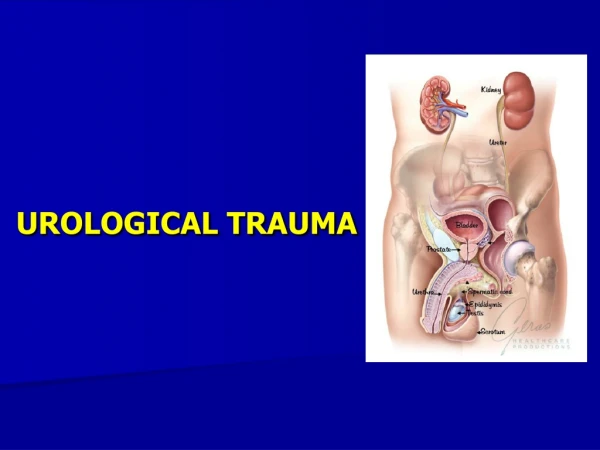
Trauma - Renal • Blunt vs penetrating trauma Blunt trauma most common with high velocity MVA Low velocity trauma – more commonly underlying abnormal kidney Penetrating injury generally stab wounds and gunshot • Blunt trauma Surgical exploration uncommon 2.6% of 913 cases in San Francisco Increasing use of radiological embolization and urological stents • Penetrating trauma Commonly require exploration 42% stab wounds explored 76% of gunshot wounds explored
Renal Trauma - Presentation • Obvious trauma – assess trauma potential • Frank haematuria common 80% to 94% of cases (but not always) N.B. especially renal pedicle injury in children and young adults deceleration injury with no haematuria Penetrating trauma – poor correlation of degree of haematuria and severity of injury • Hypotension early may be associated with loss from other injuries deceptive absence of hypotension in children
Renal Trauma - Imaging • CT scan – multi phase – non contrast, contrast arterial and venous phases and pyelographicphase. If haemodynamically stable Note importance of contrast study in assessing that pedicle intact • Intra-operative one shot IVP When patient haemodynamically unstable and emergency surgery necessary, this allows assessment of pedicle integrity in presence of identifiable non expanding peri-nephrichaematoma • Follow up imaging – pending initial staging – especially urinoma development in stage IV injury at 48 hours
Renal Trauma - Surgery • Absolute indications Severe blood loss with haemodynamic instability, not suitable for embolization Renal pedicle avulsion ? Time limits Ureteric avulsion • Relative indications Nonviable tissue – if large segments of ischaemic tissue ? % vs risk of delayed haemorrage Urinary extravasation Calyceal injury vsureteric avulsion JJ stenting with radiological drainage perc drain
Renal Trauma – Secondary Haemorrhage • 2 – 36 days post injury • Most often arteriovenous fistula or pseudo-aneurysm • 13% - 25% with grade III and IV injuries • Rx most commonly selective embolization currently
Renal Trauma - Hypertension • Incidence 0.3% - 0.9% Earliest 37 days, but up to decades after injury Average 34 months • Etiology – more likely in more severe injuries grade IV Page kidney parenchymal compression by fibrosis Renal artery stenosis, post intimal injury Arterio-venous fistulas • Diagnosis Regular blood pressure monitoring in high grade injuries ? 6 monthly lifelong
Trauma - Bladder • Uncommon • Etiology Iatrogenic most common – urology/gynaecology Spontaneous – rare in abnormal bladders eg clam cystoplasty Intoxicated – alcohol abuse with fall onto full bladder present with pain, unable to void or haematuria Traumatic – blunt trauma with high velocity MVA strong association with pelvic fracture (85% of ruptures) • Classification – ExtraperitonealvsIntraperitoneal (10% combination) • Imaging diagnosis CT delayed post contrast phase in major trauma Cystogram in iatrogenic/spontaneous/intoxicated groups
Bladder Trauma Treatment • Extraperitonealrupture Urethral catheter drainage (18F catheter) Duration pending mechanism and severity of injury 5 days up to 3 weeks Repeat cystogram prior to catheter removal • Intraperitonealrupture Traditionally surgical repair More recently, conservative, with catheter drainage and follow up cystogram
“Anterior” Urethral Trauma • Etiology Most commonly iatrogenic – forced catheterization with stricture “Fall astride” injury • History History of trauma, blood at urethral meatus and urine retention • Examination Blood at urethral meatus • Investigation Usually nil for iatrogenic Urethrogram for fall astride injuries • Treatment Nil if voiding OK for iatrogenic injury and catheter not required Cystoscopy/ endourological management in some cases Surgical repair of fall astride anterior urethral injury for complete rupture relatively easy surgery, catheter 3 weeks post op
“Posterior” Urethral Trauma • Etiology Most commonly iatrogenic – no Rx or endourological/ catheterization Associated significant pelvic injury – high level trauma potential • Clinical findings Blood at meatus + urine retention • Investigation Urethrogram +/- CT abdomen & pelvis • Management – surgical Timing pending other injuries suprapubic catheterization common Endourologicalre-alignment (flexi scope via suprapubic tract and optical urethrotome up urethra, with passage “glide” wire) Significant incidence of subsequent stricture – role of self dilatn Membranous urethroplasty – advanced surgical technique