
We present a case of a 7-year-old Albanian girl with abdominal pain and a palpable mass diagnosed with advanced ovarian dysgerminoma. Initial symptoms included generalized abdominal pain and distension, which localized to the right side over two months. Imaging revealed a large mass connected to the right ovary, leading to a total hysterectomy with bilateral salpingo-oophorectomy. Pathological examination confirmed dysgerminoma (FIGO Stage III-c). Post-surgery, she underwent chemotherapy and radiotherapy, resulting in complete remission after one year. This case emphasizes the need for thorough clinical examination and the consideration of dysgerminomas in children.

E N D
Presentation Transcript
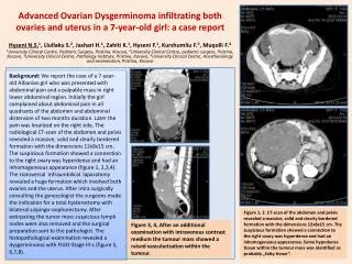
Background: We report the case of a 7-year-old Albanian girl who was presented with abdominal pain and a palpable mass in right lower abdominal region. Initially the girl complained about abdominal pain in all quadrants of the abdomen and abdominal distension of two months duration. Later the pain was localized on the right side. The radiological CT-scan of the abdomen and pelvis revealed a massive, solid and clearly bordered formation with the dimensions 12x8x15 cm. The suspicious formation showed a connection to the right ovary was hyperdense and had an inhomogeneous appearance (figure 1, 2,3,4). The transversal infraumbilicallaparatomyrevealed a huge formation which involved both ovaries and the uterus. After intra surgically consulting the gynecologist the surgeons made the indication for a total hysterectomy with bilateral salpingo-oophorectomy. After extirpating the tumor mass suspicious lymph nodes were also removed and the surgical preparation sent to the pathologist. The histopathological examination revealed a dysgerminoma with FIGO-Stage III-c (figure 5, 6,7,8). Advanced Ovarian Dysgerminoma infiltrating both ovaries and uterus in a 7-year-old girl: a case report Hyseni N.S.1, Llullaku S.2, Jashari H.1, Zahiti K.1, Hyseni F.1, Kurshumliu F.3, MuqolliF.4 1University Clinical Centre, Pediatric Surgery, Pristina, Kosovo, 2University Clinical Centre, pediatric surgery, Pristina, Kosovo, 3University Clinical Centre, Pathology Institute, Pristina, Kosovo, 4University Clinical Centre, Anesthesiology and reanimation, Pristina, Kosovo Figure 1, 2. CT-scan of the abdomen and pelvis revealed a massive, solid and clearly bordered formation with the dimensions 12x8x15 cm. The suspicious formation showed a connection to the right ovary was hyperdense and had an inhomogeneous appearance. Some hypodense tissue within the tumour mass was identified as probably „fatty tissue“. Figure 3, 4, After an additional examination with intravenous contrast medium the tumour mass showed a raised vascularization within the tumour.
Figure 7. Higher magnification showing focal prominent nucleoli of the tumor cells (x20; H&E stain). Figure 6. Medium sized tumor cells with eosinophillic cytoplasm and central nuclei with vesicular chromatin (x10; H&E stain). Figure 5.Uniform tumor cells arranged in nests, separated by delicate fibrous stroma rich in lymphocytes (×5; H&E stain) Results: We used also 4 chemotherapy-cycles (including cisplatin, etoposide, bleomycin )and radiotherapy. She is still in complete remission approximately one year after presentation • Conclusions: Our case show that inconspicuous symptoms like abdominal pain, abdominal enlargement, nausea and vomiting might occur due to a malignant ovarian germ cell tumour. • Although dysgerminomas rarely occur the physician should not exclude the possibility of dysgerminomas appearing in childhood. Therefore a complete clinical examination with radiological scans is necessary in order not to miss growing malignant formations. Adjuvant chemotherapy and radiotherapy show favourable outcome Figure 8.Lymph node metastasis with area of the tumor cells partially replacing the lymph node structure (x2.5; H&E stain).