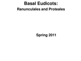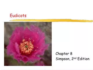
Eudicots
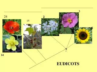
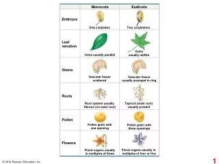
Monocots. Eudicots. Embryos. One cotyledon. Two cotyledons. Leaf venation. Veins usually netlike. Veins usually parallel. Stems. Vascular tissue scattered. Vascular tissue usually arranged in ring. Roots. Root system usually fibrous (no main root). Taproot (main root)

Eudicots
E N D
Presentation Transcript
Monocots Eudicots Embryos One cotyledon Two cotyledons Leaf venation Veins usually netlike Veins usually parallel Stems Vascular tissue scattered Vascular tissue usually arranged in ring Roots Root system usually fibrous (no main root) Taproot (main root) usually present Pollen Pollen grain with one opening Pollen grain with three openings Flowers Floral organs usually in multiples of four or five Floral organs usually in multiples of three
Reproductive shoot (flower) Apical bud Node Internode Apical bud Shoot system Vegetative shoot Blade Leaf Petiole Axillary bud Stem Taproot Lateral (branch) roots Root system
Storage roots Pneumatophores “Strangling” aerial roots
Stolon Rhizome Root Rhizomes Stolons Tubers
Spines Tendrils Storage leaves Stem Storage leaves Reproductive leaves
Dermal tissue Ground tissue Vascular tissue
Parenchyma cells with chloroplasts (in Elodea leaf) (LM) 60 m
Collenchyma cells (in Helianthus stem) (LM) 5 m
5 m Sclereid cells (in pear) (LM) 25 m Cell wall Fiber cells (cross section from ash tree) (LM)
100 m Vessel Tracheids Pits Tracheids and vessels (colorized SEM) Perforation plate Vessel element Vessel elements, with perforated end walls Tracheids
Sieve-tube elements: longitudinal view (LM) 3 m Sieve plate Sieve-tube element (left) and companion cell: cross section (TEM) Companion cells Sieve-tube elements Plasmodesma Sieve plate 30 m Nucleus of companion cell 15 m Sieve-tube elements: longitudinal view Sieve plate with pores (LM)
Primary growth in stems Epidermis Cortex Primary phloem Shoot tip (shoot apical meristem and young leaves) Primary xylem Pith Vascular cambium Secondary growth in stems Lateral meristems Cork cambium Cork cambium Axillary bud meristem Periderm Pith Cortex Primary phloem Secondary phloem Primary xylem Root apical meristems Secondary xylem Vascular cambium
Apical bud Bud scale Axillary buds This year’s growth (one year old) Leaf scar Bud scar Node One-year-old branch formed from axillary bud near shoot tip Internode Last year’s growth (two years old) Leaf scar Stem Bud scar Growth of two years ago (three years old) Leaf scar
GLABRA-2 is expressed, and the cell remains hairless. Cortical cells GLABRA-2 is not expressed, and the cell will develop a root hair. 20 m The root cap cells will be sloughed off before root hairs emerge.
Cortex Vascular cylinder Dermal Ground Epidermis Vascular Zone of differentiation Root hair Zone of elongation Mitotic cells Zone of cell division (including root apical meristem) 100 m Root cap
Epidermis Cortex Endodermis Vascular cylinder Pericycle Core of parenchyma cells Xylem 100 m Phloem 100 m (a) Root with xylem and phloem in the center (typical of eudicots) (b) Root with parenchyma in the center (typical of monocots) Endodermis Pericycle Xylem Phloem Dermal Ground Vascular 70 m
Epidermis Emerging lateral root 100 m Lateral root Cortex Pericycle Vascular cylinder
Shoot apical meristem Leaf primordia Young leaf Developing vascular strand Axillary bud meristems 0.25 mm
Guard cells Stomatal pore 50 m Dermal Epidermal cell Ground Sclerenchyma fibers Cuticle Vascular Stoma (b) Surface view of a spiderwort (Tradescantia) leaf (LM) Upper epidermis Palisade mesophyll Spongy mesophyll Lower epidermis 100 m Bundle- sheath cell Cuticle Xylem Vein Phloem Guard cells Air spaces Guard cells Vein (a) Cutaway drawing of leaf tissues (c) Cross section of a lilac (Syringa) leaf (LM)
Phloem Xylem Sclerenchyma (fiber cells) Ground tissue Ground tissue connecting pith to cortex Epidermis Pith Dermal Cortex Ground Epidermis Vascular bundles Vascular bundle Vascular 1 mm 1 mm (b) Cross section of stem with scattered vascular bundles (typical of monocots) (a) Cross section of stem with vascular bundles forming a ring (typical of eudicots)
Pith (a) Primary and secondary growth in a two-year-old woody stem Primary xylem Vascular cambium Primary phloem Epidermis Cortex Cortex Epidermis Primary phloem Vascular cambium Growth Vascular ray Periderm Secondary phloem Cork cambium Primary xylem Vascular cambium Cork Bark Pith Secondary xylem Late wood Secondary xylem Early wood Secondary phloem Cork First cork cambium 1 mm Periderm (mainly cork cambia and cork) Growth Vascular ray Growth ring 1.4 mm (b) Cross section of a three-year- old Tilia (linden) stem (LM) Secondary phloem Most recent cork cambium Layers of periderm Secondary xylem Bark Cork
Vascular cambium Growth Vascular cambium Secondary phloem Secondary xylem After one year of growth After two years of growth
Growth ring Vascular ray Heartwood Secondary xylem Sapwood Vascular cambium Secondary phloem Bark Layers of periderm