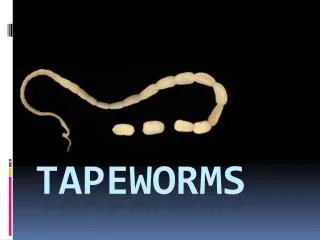
Cestoda (Tapeworms)
Cestoda (Tapeworms). Taenia saginata Taenia solium Echinococcus granulosus. Cestoda (Tapeworms). General characteristics * Parasitize in the small intestine of humans.

Cestoda (Tapeworms)
E N D
Presentation Transcript
Cestoda (Tapeworms) Taenia saginata Taenia solium Echinococcus granulosus
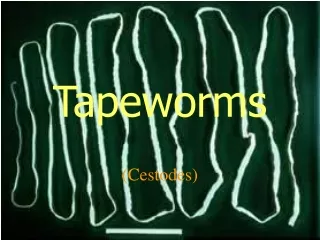
Cestoda (Tapeworms) • General characteristics *Parasitize in the small intestine of humans. *They are hermaphrodites and consist of the head (scolex), followed by an unsegmented germinative section (neck) and a posterior chain of segments (proglottids). *There are no digestive organs, so nutrients are taken up through the absorptive integument.
Cestoda (Tapeworms) *The life cycle of cestodes include one or two intermediate hosts. *Humans can also be infected by larval stages of various tapeworm species (cysticerci, metacestodes). *These stages develop in body tissues and generally cause considerably greater pathological damage than the intestinal cestode stages.
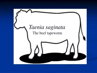
Taenia speciesCausative agents of taeniosis and cysticercosis *Taeniosis is a small intestine infection of humans caused by Taenia species. *In T. saginata, the intermediate hosts are cattle. *In the musculature of the cattle cysticerci develop and can be ingested by humans who eat raw beef. * The cysticerci of T. solium develop in the musculature of pigs.
Taenia saginata (Beef Tapeworm)Causative agent ofT. saginata taeniosis • Occurrence. This species occurs worldwide the number of infected humans is estimated to be between 40 and 60 million. The parasite • *T.saginata grows as long as 10m and has a scolex with four suckers. • *Proglottids at the posterior end of the chain are longer than wide and each contains a treelike branched uterus containing 80 000–100 000 eggs.
Taenia saginata (Beef Tapeworm)Causative agent ofT. saginata taeniosis *The eggs are released when a proglottid detaches from the tapeworm in the intestinal lumen or when a segment disintegrates • outside the host. • *The eggs are small diameter 30–40 µm) and round. • *The eggs are highly resistant and can remain infective in a moist environment for weeks or • months.
Taenia saginata (Beef Tapeworm)Causative agent ofT. saginata taeniosis • Life cycle • * Carried by feces of humans infected with Taenia, they contaminate pastures or feed either directly or via sewage. • * When the cattle ingest the eggs, the oncospheres hatch in the small intestine, migrate into the intestinal wall, and are transported with the bloodstream into the striated musculature of the cattle .
Taenia saginata (Beef Tapeworm)Causative agent ofT. saginata taeniosis *Into the striated musculature they develop into the infective metacestodes or cysticerci(= Cysticercus bovis) within three to four months. *Each cysticercus is a pea-sized, fluid-filled cyst containing a single invaginated scolex *Humans are infected by ingesting raw or undercooked beef containing cysticerci.
Taenia saginata (Beef Tapeworm)Causative agent ofT. saginata taeniosis *In the small intestine, the cysticercus evaginates the scolex. *The scolex attaches to the mucosa of the upper small intestine, and develops into an adult tapeworm, which can live for years or even decades. * About two to three months after the infection, the first gravid segments detach from the strobila • and then appear in feces or they can migrate out of the intestine without defecation. *The segments remain motile for some time and frequently leave the stools.
Taenia saginata (Beef Tapeworm)Causative agent ofT. saginata taeniosis • Pathogenesis and clinical manifestations. • *T.saginata causes morphological changes like villus deformation, enterocyte proliferation,cellular mucosal infiltration, and functional disturbances. • * Eosinophilia may occur sometimes. • *The infection takes an asymptomatic course in about 25% of • cases. • * Symptoms of infection include nausea, vomiting, upper abdominal pains, diarrhea or constipation and increased or decreased appetite. • Infection does not confer levels of immunity sufficient to prevent reinfection.
Taenia saginata (Beef Tapeworm)Causative agent ofT. saginata taeniosis Therapy and prevention. * The drug of choice is the highly effective praziquantel. * Albendazole, mebendazole, and paromomycin are less reliable. * Meat containing numerous cysticercihas to be confiscated, but meat with small numbers of * Cysticerci can be used for human consumption after deep-freezing that is lethal to the parasites. * Individual prophylaxis consists of not eating beef that is raw or has not been deep-frozen.
Taenia saginata (Beef Tapeworm)Causative agent ofT. saginata taeniosis • Diagnosis. • * A Taenia infection is easy to diagnose if the 1.5–2cm long and 0.7cm wide segments are eliminated in stool. • * Morphological species differentiation is often not possible based on the gravid proglottids, but can be done by DNA-analysis (PCR). • * T. Saginata eggs are shed irregularly in stool and cannot be differentiated morphologically from T. solium eggs. • * Using an ELISA, coproantigens are detectable in stool fluid even when neither proglottids nor eggs are being excreted.
Echinococcus granulosus • * The most important species of the genus Echinococcus are Echinococcus granulosus and E. multilocularis parasite of fox species, dogs, cats, and other carnivores). • * Both species occur in Europe. • * Their metacestodes can cause cystic echinococcosis (CE, hydatid disease) or alveolar echinococcosis (AE) in humans.
Echinococcus granulosus • * Humans are infected by peroral ingestion of Echinococcus eggs, from which in CE, liquid-filled cystic metacestodes (the hydatids) develop, particularly in the liver and lungs. • * The metacestodes primarily parasitize the liver, where they proliferate like a tumor and form small cysts; secondary metastatic spread to other organs is possible.
Echinococcus granulosus Echinococcus species are small tapeworms that parasitize the small intestine of carnivores and produce eggs that are shed to the environment by the host. Pathogenic larval stages (metacestodes) develop following peroral ingestion of such eggs by the natural intermediate hosts (various mammalian species), as well as in humans and other accidental hosts (which do not play a role in the life cycle).
Echinococcus granulosus Morphology and development Adult stage. E. granulosus is a 4–7mmlong tapeworm with a scolex and normally three (two to six) proglottids. A notable characteristic is the uterus with its lateral sacculations, containing up to • 1500 eggs
Echinococcus granulosus • Definitive (final) and intermediate hosts. • The definitive host for E. granulosus is the dog, whereby other Canidae (jackal, dingo, and • other wild canids) are involved in certain regions. • Herbivorous and omnivorous vertebrates function as intermediate hosts, in particular domestic animals (ruminants, pigs, horses, camels) .
Echinococcus granulosus • Life cycle The adult tapeworms live in the small intestine of the definitive host for about six months, a few for up to two years. • Eggs are either released from gravid proglottids in the intestine and shed with feces or pass out of the host still enclosed in the tapeworm segments. • The eggs (diameter approx. 30–40 µm) are nearly spherical, contain an oncosphere.
Echinococcus granulosus • They cannot be morphologically differentiated from the eggs of other Echinococcus or Taenia species • Infection of the intermediate hosts, humans, and other accidental hosts is by peroral ingestion of eggs, from which the oncospheres are released in the small intestine, penetrate into its wall and migrate hematogenously into the liver, as well as sometimes into the lungs and other organs. • At first, the oncospheres develop into little vesicles, then gradually into metacestodes.
Echinococcus granulosus • The metacestode of E. granulosus (also known as hydatid cysts, is normally a fluid-filled cyst with one or multiple chambers, thewall of which is made up of an inner, cellular, germinative layer and an outer, acellular, laminated layer (cuticular layer), enclosed by a layer of host connective tissue
Echinococcus granulosus Brood capsules develop five to six months or later on the germinative layer, each containing up to 20 or more protoscoleces with four suckers and tow rows of rostellar hooks. The thin brood capsules burst to release free protoscoleces into the hydatid fluid, which form, together with the brood capsules, their remains and calcareous corpuscles the so called “hydatid sand.” Cysts in humans often contain smaller daughter cysts.
Echinococcus granulosus • The life cycle is completed when carnivores ingest E. granulosus cysts containing mature protoscoleces. • It develop in the small intestine of the definitive hosts within five to eight weeks. • Acute symptoms may appear following spontaneous, traumatic, or intraoperative cyst ruptures, whereby the release of antigen containing hydatid fluid can cause • symptoms of anaphylactic shock. • There is also a risk that protoscoleces will be • released and develop into new cysts in the human host On • the other hand, cyst rupture can also result in spontaneous cure.
Echinococcus granulosus • Diagnosis is based on detection of cysts using imaging techniques (ultrasonography,computer tomography, thoracic radiography, etc.) in connection • with serological antibody detection Specific antibodies • occur in about 90–100% of patients with cystic hepatic echinococcosis, but in only about 60–80% of cases with pulmonary echinococcosis. • Diagnostic cyst puncture is generally not advisable due to the risks described above (secondary echinococcosis, anaphylactic reactions).
Echinococcus granulosus • The disease can be cured by removing the Echinococcus cysts surgically.Inoperable patients can be treated during several months with albendazole or mebendazole. • Chemotherapy results in cure in about 30% of cases and in improvement in afurther 30–50% (WHO, 1996). PAIR (puncture aspiration injection reaspiration) Therapy is a new technique still under evaluation: after puncturing the cysts (not all cysts are suitable, e.g., pulmonary cysts!) under ultrasonic Guidance, most of the hydatid fluid is aspirated, after which an adequate amount of 95% ethanol is injected into the cyst, left in it for 15 minutes and removed (reaspirated).
Echinococcus granulosus • Control and prevention. Control of CE in humans includes regular mass treatment of dogs to eliminate E. granulosus, preventing access of dogs to viscera of domestic or wild animals, and dog population control. • Special hygienic principles must be observed when handling dogs in endemic areas. • -------------------------------