Download
1 / 67
670 likes | 818 Vues
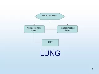
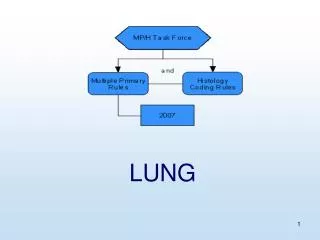
LUNG. Equivalent Terms, Def, Charts, Tables, Illustrations. Equivalent Terms. Default multiple tumors with only one biopsied Equivalent Neuroendocrine ca – carcinoid. Table 1 Instructions.

E N D
Equivalent Terms • Default • multiple tumors with only one biopsied • Equivalent • Neuroendocrine ca – carcinoid
Table 1 Instructions Use this table to select combination/mixed histology codes. Compare the terms in the diagnosis to the terms in columns 1 and 2. If the terms match, abstract the case using the ICD-O-3 histology code in column 4. Use the combination/mixed codes listed in this table only when the histologies in the tumor match the histologies listed below. Use the combination/mixed codes for a single tumor when all histologies are present in a single tumor. Note: This table is not a complete listing of histologies that may occur in the lung
M1 When it is not possible to determine if there is a single tumor ormultiple tumors, opt for a single tumor and abstract as a singleprimary.
M1 Notes Note 1: Use this rule only after all information sources have been exhausted. Note 2: Use this rule when only one tumor is biopsied but the patient has two or more tumors in one lung and may have one or more tumors in the contralateral lung. (See detailed explanation in Lung Equivalent Terms and Definitions).
M2 A single tumor is always a singleprimary. Note: The tumor may overlap onto or extend into adjacent/contiguous site or subsite.
M3 Tumors in sites with ICD-O-3 topography codes that are different at the second (Cxxx) and/or third character (Cxxx) are multiple primaries. Note: This is a change in rules; tumors in the trachea (C33) and in the lung (C34) were asingle lung primary in the previous rules.
M4 At least one tumor that is non-small cell carcinoma (8046) and another tumor that is small cell carcinoma (8041-8045) are multiple primaries.
M5 A tumor that is adenocarcinoma with mixed subtypes (8255) and another that is bronchioloalveolar (8250-8254) are multiple primaries.
M6 A single tumor in eachlung is multiple primaries.
M6 Note When there is a single tumor in each lung abstract as multiple primaries unless stated or proven to be metastatic.
M7 Multiple tumorsin both lungs with ICD-O-3 histology codes that are different at the first (xxxx), second (xxxx) or third (xxxx) number are multiple primaries.
M8 Tumors diagnosed more than three (3) years apart are multiple primaries.
M9 An invasive tumor following an in situ tumor more than 60 days after diagnosis is a multiple primary.
M9 Notes Note 1: The purpose of this rule is to ensure that the case is counted as an incident (invasive) case when incidence data are analyzed. Note 2: Abstract as multiple primaries even if the medical record/physician states it is recurrence or progression of disease.
M10 Tumors with non-small cell carcinoma, NOS (8046) and a more specific non-small cell carcinoma type (Chart 1) are a single primary.
M11 Tumors with ICD-O-3 histology codes that are different at the first (xxxx), second (xxxx) or third (xxxx) number are multiple primaries.
M11 Note Note: Adenocarcinoma in one tumor and squamous cell carcinoma in another tumor are multiple primaries.
M12 Tumors that do not meet any of the above criteria are a single primary.
M12 Notes Note 1: When an invasive tumor follows an in situ tumor within 60 days, abstract as a single primary. Note 2: All cases covered by this rule are the same histology.
M12 Examples The following are examples of cases that use Rule M12. This is NOT intended to be an exhaustive set of examples; there are other cases that may be classified as a single primary.
M12 Examples Warning: Using only these case examples to determine the number of primaries can result in major errors.
H1 Code the histology documented by the physician when there is no pathology/cytology specimen or the pathology/cytology report is not available.
H1 Note 1 • Note 1: Priority for using documents to code the histology • Documentation in the medical record that refers to pathologic or cytologic findings • Physician’s reference to type of cancer (histology) in the medical record • CT, PET, or MRI scans • Chest x-rays
H1 Notes 2 and 3 Note 2: Code the specific histology when documented. Note 3: Code the histology to 8000 (cancer/malignant neoplasm, NOS) or 8010 (carcinoma, NOS) as stated by the physician when nothing more specific is documented.
H2 Code the histology from a metastatic site when there is no pathology/cytology specimen from the primary site. Note: Code the behavior /3
H3 Code the histology when only one histologic type is identified. Note:Do not code terms that do not appear in the histology description.
H3 Examples Example 1: Do not code squamous cell carcinoma non-keratinizing unless the words “non-keratinizing” actually appear in the diagnosis. Example 2: Do not code bronchioalveolar non-mucinous unless the words “non-mucinous” actually appear in the diagnosis.
H4 Code the invasive histologic type when a single tumor has invasive and in situ components.
H5 Code the most specific term using Chart 1 when there are multiple histologies within the same branch.
H5 Continued Examples of histologies within the same branch are • Cancer/malignant neoplasm, NOS (8000) and a more specific histology or • Carcinoma, NOS (8010) and a more specific carcinoma or • Adenocarcinoma, NOS (8140) and a more specific adenocarcinoma or • Squamous cell carcinoma, NOS (8070) and a more specific squamous cell carcinoma or • Sarcoma, NOS (8800) and a more specific sarcoma
H5 Note The specific histology may be identified as type, subtype, predominantly, with features of, major, or with ____differentiation
H5 Examples Example 1: Adenocarcinoma, predominantly mucinous. Code 8480 (mucinous adenocarcinoma). Example 2: Non-small cell carcinoma, papillary squamous cell. Code 8052 (papillary squamous cell carcinoma).
H6 Code the appropriate combination/mixed code (Table 1) when there are multiple specific histologies or when there is a non-specific with multiple specific histologies.
H6 Note and Examples Note: The specific histologies may be identified as type, subtype, predominantly, with features of, major, or with ____differentiation. Example 1 (multiple specific histologies):Solid and papillary adenocarcinoma. Code 8255 (adenocarcinoma with mixed subtypes).
H6 Examples continued Example 2 (multiple specific histologies): Combined small cell and squamous cell carcinoma. Code 8045 (combined small cell carcinoma). Example 3 (non-specific with multiple specific histologies): Adenocarcinoma with papillary and clear cell features. Code 8255 (adenocarcinoma with mixed subtypes).