The Structure of the Cell Membrane
190 likes | 502 Vues
The Structure of the Cell Membrane. A soap molecule has a long hydrocarbon chain and a charged head. Soap will form a mono-molecular film on water. Agitating soapy water makes micelles. Soap bubbles are made from a double layer of soap molecules sandwiching a thin layer of water.

The Structure of the Cell Membrane
E N D
Presentation Transcript
A soap molecule has a long hydrocarbon chain and a charged head. Soap will form a mono-molecular film on water.
Soap bubbles are made from a double layer of soap molecules sandwiching a thin layer of water.
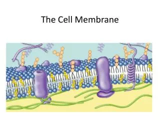
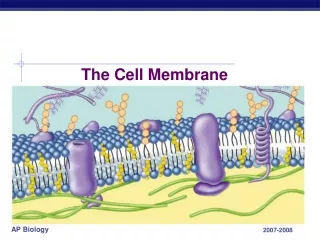
Phospholipids, the main component of cell membranes, behave like soap molecules.
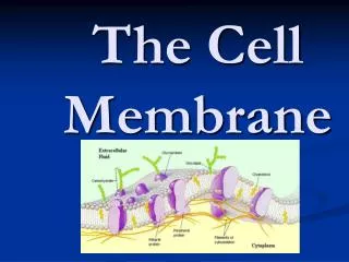
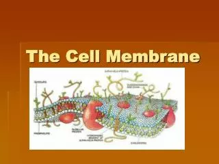
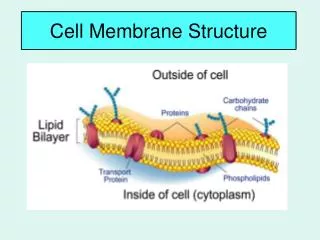
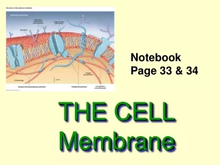
Introduction • A thin outer layer called the cell membrane surrounds all cells. • This membrane has four main functions: • allow the transport of raw materials into the cell • allow the transport of manufactured products and wastes out of the cell • prevent the entry of unwanted material into the cell • prevent the leakage of essential matter out of the cell
Introduction • Cellular functions depend on a watery environment like the one that is found on the inside of the cell. • In order to maintain their integrity, cells need to be surrounded by an environment through which water cannot flow. A membrane composed of fatty molecules serves this purpose.
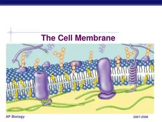
History of the Cell Membrane • In 1924, using an electron microscope, two Dutch physicians, E. Gorter and F. Grendel found that the cell membrane was composed primarily of phospholipids (shown on the right).
History of the Cell Membrane • They deduced, based on the properties of phospholipids, that the cell membrane was in fact a bilayer.
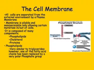
History of the Cell Membrane • Cell membranes form because these phospholipid molecules spontaneously orient themselves in a double layer having a fatty inside and a water soluble outside.
History of the Cell Membrane • Phospholipids are made up of a glycerol backbone with a hydrophilic head region containing a phosphate group and a hydrophobic tail region containing a saturated fatty acid and an unsaturated fatty acid. • The fact that it has both types of fatty acids ensures the cell membrane is fluid.
History of the Cell Membrane • By the 1930s experimental evidence showed that proteins were also part of the cell membrane. • In 1935, James Danielli and Hugh Davson proposed the sandwich model: a phospholipid bilayer between two layers of protein with pores for molecules to travel through.
History of the Cell Membrane • Stronger electron microscopes would show that the cell membrane was not covered in protein, but rather had protein embedded in it.
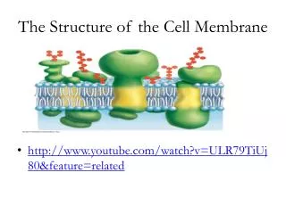
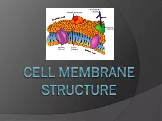
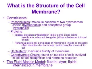
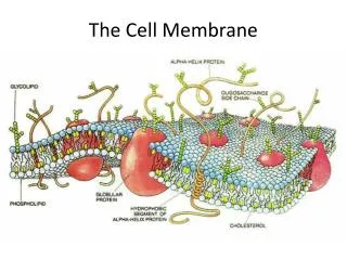
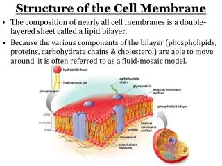
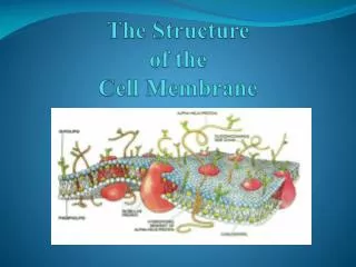
History of the Cell Membrane • In 1972, S. J. Singer and Garth Nicholson presented the fluid mosaic model of the cell membrane, which displayed the cell membrane as an integration of proteins and other molecules into the phospholipid bilayer.
Structures in the Fluid Mosaic Model of the Cell Membrane • Cholesterol is interspersed throughout the cell membrane to add rigidity to it. • It also allows the cell membrane to stay fluid over a wider range of temperatures.
Structures in the Fluid Mosaic Model of the Cell Membrane • Various proteins are associated with the cell membrane. • Integral Proteins (a.k.a. trans-membrane proteins) span the width of the cell membrane and create channels through which charged molecules or large molecules can pass through. • Peripheral Proteins are found on the surface of the cell membrane and are primarily used in cell to cell signaling with surface carbohydrate chains or linking with the cytoskeleton for support.
Structures in the Fluid Mosaic Model of the Cell Membrane • The cytoskeleton is attached to the cell membrane for added stability, since membrane proteins and phospholipids can shift places in the membrane.