Download
1 / 33
360 likes | 1.17k Vues
CT Urography and applications in uroephithelial tumors. Orith Portnoy Dept. of Diagnostic Radiology Sheba Medical Center, Sakler School of Med. Tel-Aviv University, Israel. IVP (intravenous pyelography).

E N D
CT Urography and applications in uroephithelial tumors Orith Portnoy Dept. of Diagnostic Radiology Sheba Medical Center, Sakler School of Med. Tel-Aviv University, Israel
IVP (intravenous pyelography) • Initial modality for upper tract imaging in hematuria, flank pain & others for 7 decades. • Less sensitive than CT for: • Renal masses (21% for 2 cm mass) • Urinary tract stones • Renal inflammation • Renal trauma
CT Urography (CTU) • Single detector MDCT volumetric acquisition high resolution reconstructions • Both renal parenchyma and urothelium shown in a single examination • Shortening schedule for diagnostic evaluation (hematuria)
CTU at Sheba • Since 6/2004 • ~ 500 studies • GE MDCT 16/64 slice, Philips MDCT BR 40/64 slice
ProtocolCTU • Monitored by a radiologist • Non contrast phase (low dose) • Nephrographic phase (100s delay) saline IV • Excretory phase (400-800 slices) tailored • Reconstructions on a 4.1 or 4.2 GE workstation
CTU – Rec. bladder TCC 80 Y.O. man Macrohematuria S/P 17 operations for bladder TCC
56 Y.O. man macrohematuria Rec. bladder TCC seen at cystoscopy Posterior view
CTU and US 46 Y.O. women 1 event of macrohematuria
CTU and IVP • 68 Y.O. man • Left flank pain US (stone) lithothripsy hematuria post 3w IVPcystoscopy (susp. tumor)
61 Y.O. man Recurrent macrohematuria 6 mo. before – US, IVP, cystoscopy
CTU and PET CT Bladder TCC and CLL Retrograde pyelography – narrowed ureter
Sensitivity • Detection of upper tract urothelial tumors by CTU – 91-94% in relation to biopsy (Dillman AbdImaging 2008) • Detection of bladder tumors: microhematuria – 40% vs. cystoscopy, macrohematuria high risk – 93% sens., 99% spec.(Albani J Urol 2007, Turney BJU 2006) • High risk: >40y, macrohematuria, smoking, GU tumor P/H, occupational exposure
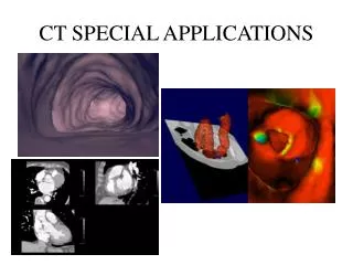
Types of Urinary Diversion after Cystectomy • Incontinent diversion (ileal, colonic) • Continent cutaneous catheterizable reservoir • Orthotopic neobladder
Imaging after bladder reconstruction • Complications • Recurrence • Understanding the reconstruction anatomy helps diagnose complications • US, IVP, cystography/lupographyantegrade/retrograde pyelography,CT,nuclear medicine • CT-UROGRAPHY
68 Y.O. man • 6 years post bladder replacement d/t TCC • 6 months intermittent macrohematuria
CTU - Disadvantages • Radiation dose • Mean effective dose: 23-35 mSv • CTU 1.5 more than standard IVP Nawfel et alRadiology 2004 • Time consuming processing, reviewing • Lack large scale research on cost-effectiveness
CTU - summary • Useful diagnostic examination that allows comprehensive evaluation of urinary tracts • Problem solving tool with other modalities • Becoming the primary imaging study for the work-up of patients with hematuria and other genitourinary conditions • Shorter diagnostic evaluation, decrease need for ureteroscopies • Tailored examination can save radiation • Referrals should be limited (urologists)
CTU and “regular” CT 66 Y.O. man 1 year post partial nephrectomy for RCC. New hydronephrosis on CT, suspect rec. obstructing tumor. POST. VIEW