Download
1 / 36
380 likes | 528 Vues
Apoptóza a genová terapie. Mgr. Jan Bouchal, PhD. Laboratoř molekulární patologie LF UP Olomouc http://lmp.upol.cz, http://ustavpatologie.upol.cz. Funkce nádorového supresoru p53. Cell options. keep working. apoptosis. fuse. stress responses. hypertrophy. shrink. enlarge & divide.

E N D
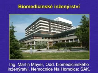
Apoptóza a genová terapie Mgr. Jan Bouchal, PhD. Laboratoř molekulární patologie LF UP Olomouc http://lmp.upol.cz, http://ustavpatologie.upol.cz
Cell options keep working apoptosis fuse stress responses hypertrophy shrink enlarge & divide de-differentiate transdifferentiate senescence RETREATORADVANCE
Apoptóza neboli programovaná buněčná smrt • Charakterizována kondenzací chromatinu, puchýřkovatěním cytoplazmatické membrány (budding) a tvorbou apoptotických tělísek • Apoptotická tělíska jsou fagocytována a nedochází k zánětlivé reakci • http://en.wikipedia.org/wiki/Apoptosis • http://www.celldeath.de/mainfram.htm
Incomplete differentiation in two toes (syndactyly) due to lack of apoptosis
WHERE can APOPTOSIS be ENCOUNTERED ? ... Growth of Embryo ... Tissue Homeostasis ... Immunology ... Chronic viral diseases ... Neurodegenerative diseases ... Reperfusion injury ... Insuline-dependent Diabetes ... Atherosclerosis ... Myokard Infarction ... AIDS ... Development and Treatment of Malignancies
THE APOPTOTIC PATHWAY Triggers Modulators Effectors Substrates DEATH . Many cellular proteins . DNA . FADD . TRADD . FLIP . Bcl-2 family . Cytochrome c . p53 . Mdm2 . Caspases . Growth factor Deprivation . Hypoxia . Loss of adhesion . Death receptors . Radiation . Chemotherapy
Receptorová a mitochondriálnídráha apoptózy Caspázy (cysteinyl aspartate specific proteinases; neaktivní prekurzory -procaspázy) Receptory smrti Molekuly v mezimemránovém prostoru mitochondrií (nejasné teorie – jednou z nich je tvorba pórů)
WHERE can APOPTOSIS be ENCOUNTERED ? ... Growth of Embryo ... Tissue Homeostasis ... Immunology ... Chronic viral diseases ... Neurodegenerative diseases ... Reperfusion injury ... Insuline-dependent Diabetes ... Atherosclerosis ... Myokard Infarction ... AIDS ... Development and Treatment of Malignancies
Metody detekce apoptózy („alespoň dvě“) Světelná, fluorescenční a elektronová mikroskopie (morfologie) • kondenzace chromatinu, puchýřkovatění membrány (budding), apoptotická tělíska • další znaky (viz níže) Substráty caspáz • Imunohistochemická detekce štěpeníPARP, laminu B, keratinu 18, transglutaminázy Mitochondriální funkce • změny membránového potenciálu mitochondriální membrány (rhodamin 123, Mitotracker) • uvolňování cytochromu c Změny na cytoplazmatické membráně • expozice fosfatidylserinu a vazba Annexinu V • permeabilita membrány a vazba barviv na DNA (propidium iodid, Hoechst, DAPI) – odlišení od nekrózy Změny DNA • Apoptotický DNA žebřík • značení štěpených konců DNA (TUNEL, ISNTA)
Metody detekce apoptózy Světelná, fluorescenční a elektronová mikroskopie (morfologie) kondenzace chromatinu, puchýřkovatění membrány (budding), apoptotická tělíska
Metody detekce apoptózy Substráty kaspáz • detekce štěpení keratinu 18 (protilátka M30 proti štěpenému fragmentu), laminu B (inverzní průkaz)
Metody detekce apoptózy Změny na cytoplazmatické membráně - expozice fosfatidylserinu a vazba Annexinu V Změny DNA Apoptotický DNA žebřík
Další typy buněčné smrti Normální buňka Nekrotická Apoptotická Autofagická
Nekróza • Pasivní buněčná smrtv důsledku fyzikálního nebo chemického poškození • Charakteristická vakuolizace, permeabilizace cytoplazmatické membrány avyvolání místní zánětlivé odpovědi (významné během mikrobiálních infekcí)
Autofagie • Morfologické znaky podobné s nekrózou • Dochází ke strávení části vlastního buněčného materiálu (auto – fagie) • Nejasný význam • - buď další forma programované buněčné smrti • - nebo strategie pro přežití v období nedostatku energie
Hlavní znaky Apoptóza • Charakterizována kondenzací chromatinu, puchýřkovatěním cytoplazmatické membrány (budding) a tvorbou apoptotických tělísek, které jsou následně fagocytovány • Receptorová (receptory smrti, caspáza-8) a mitochondriální dráha (rodina proteinů Bcl2, cytochrom C, caspáza-9) Nekróza • Charakteristická vakuolizace, permeabilizace cytoplazmatické membrány avyvolání zánětlivé odpovědi(významné během mikrobiálních infekcí) Autofagie • Pravděpodobněstrategie pro přežitív období nedostatku energie
Genová terapie • Při zrodu genové terapie se uvažovalo především o léčbě vrozených monogenních chorob (např. ADA - Adenosine Deaminase Deficiency, cystic fibrosis, Huntington's chorea, muscular dystrophy ) • V současné době se většina úsilí orientuje na získané choroby, především zhoubné nádory • Genová terapie v léčbě nádorů • Je lehčí buňku zničit, než ji opravit (vyvolání apoptózy, …) • Zpravidla stačí krátká exprese genu (problém umlčení cizorodých genů, zpravidla metylací) • Vzhledem k povaze onemocnění jsou pacienti ochotni podstoupit nové postupy
Germ line gene therapy • In the case of germ line gene therapy, germ cells, i.e., sperm or eggs, are modified by the introduction of functional genes, which are ordinarily integrated into their genomes. This option is prohibited for application in human beings, at least for the present, for a variety of technical and ethical reasons. • Somatic cell gene therapy • In somatic cell gene therapy, the gene is introduced only in somatic cells, especially of those tissues in which expression of the concerned gene is critical for health. Expression of the introduced gene relieves/ eliminates symptoms of the disorder. • Broad methods • A normal gene may be inserted into a nonspecific location within the genome to replace a nonfunctional gene. This approach is most common. • An abnormal gene could be swapped for a normal gene through homologous recombination. • The abnormal gene could be repaired through selective reverse mutation, which returns the gene to its normal function. • The regulation (the degree to which a gene is turned on or off) of a particular gene could be altered.
Není možno zasáhnout všechny buňky • Sebevražedné geny a by-stander efekt
„…Currently gene therapy is being used to create recombinant cancer vaccines. …“ Cross et Burmester: Gene Therapy for Cancer Treatment: Past, Present and Future. Clinical Medicine and Research 2006;4:218-227.
From donated blood (usually leukapheresis) precursor cells – monocytes are extracted and in Step 1 of cell culture, transformed into dendritic cells. From tumour tissue specimens obtained from surgery, a tumour-specific antigen is produced with which dendritic cells are loaded. Additionally, dendritic cells are also loaded with a control antigen – key-hole limpet hemocyanin (KLH).The control antigen enables monitoring of the success of vaccination. After an activation step, Step 2, the ‘mature’ antigen-loaded dendritic cells are administered to the patient in the form of vaccination.
Onkolytické viry Oncolytic gene therapy vectors are generally viruses that have been genetically engineered to target and destroy cancer cells while remaining innocuous to the rest of the body. A number of different viruses have been used for this purpose, including vaccinia, adenovirus, herpes simplex virus type I, reovirus and Newcastle disease virus.38 However, there are several unique stumbling blocks for oncolytic virotherapy in humans. Most people have antibodies to the common viruses used for therapy development which often leads to an immune response that clears the viral agent before it has had time to infect cells. In addition, the use of replication competent viral particles often calls for increased safety precautions, making clinical trials more expensive and cumbersome. In a trial using a modified vaccinia virus to treat breast and prostate cancer, patients were required to be isolated in a specialized hospital facility for a week to ensure that the virus had completely cleared before being allowed back into the general population. The most notable adenoviral therapy is the ONYX-015 viral therapy. ONYX-015 is an adenovirus that has been engineered to lack the viral E1B protein. Without this protein, the virus is unable to replicate in cells with a normal p53 pathway. In addition, the E1B protein is essential for RNA export during viral replication.45 Cancer cells often have deficiencies in the p53 pathway due to mutations and thus, allow ONYX-015 to replicate and lyse the cells. Cancer cells also exhibit altered RNA export mechanisms that allow for the export of viral RNA even in the absence of the E1B protein. ONYX-015 has been tested in phase I and II trials on squamous cell carcinoma of the head and neck that resulted in tumor regression which correlated to the p53 status of the tumor. The second type of oncolytic virotherapy undergoing clinical trials uses herpes simplex virus type 1. Two vectors, G207 and NV1020, are currently in phase I and phase II trials for treatment of intractable cancers. Mutations in several genes of these herpes viruses ensure that they replicate efficiently only in cancerous cells.The lytic portion of the life cycle directly kills cells and the thymidine kinase that is expressed from the viral genes sensitizes cells to ganciclovir.
Challenges in Gene Therapy http://learn.genetics.utah.edu/units/genetherapy/gtchallenges/
From Research to Trials (15 years for ADA) STEP 1: Learn about the diseaseIs the disorder a good candidate for gene therapy? To find out, study the disease. 1) Get money for the project 2) Get approval for the project 3) Perform clinical research 4) Perform biological research 5) DECISION: Is the disorder a good candidate for gene therapy? STEP 2: Design a gene therapy 1) Use your knowledge of the disorder to design a gene therapy 2) Test the therapy in appropriate models of the disease 3) DECISION: Does your therapy look promising? http://learn.genetics.utah.edu/units/genetherapy/gtresearch/
STEP 3: Get money and approval for clinical trials 1) Get money for the trials 2) Get approval for the trials STEP 4: Phase One clinical trial 1) Establish safety and dosage limits in a small group of people (20-80) 2) DECISION: Does your therapy still look promising? STEP 5: Phase Two clinical trial 1)Test the efficacy and safety in a larger group of people (100-300) 2) DECISION: Is your therapy effective in a larger group of people? STEP 6: Phase Three clinical trial 1) Test the therapy in a large group of people (1,000-3,000) 2) DECISION: Is your treatment successful? STEP 7: Get FDA approval for general clinical use 1) Write proposals, fill out paperwork, answer questions and wait for approval STEP 8: Phase Four clinical trial 1) Further test the efficacy and optimal use of the treatment in general use
Shrnutí • Genová terapie v léčbě nádorů • Je lehčí buňku zničit, než ji opravit a zpravidla stačí krátká exprese genu • Vzhledem k povaze onemocnění jsou pacienti ochotni podstoupit nové postupy • Genová terapie (by-stander efekt) • Imunoterapie