Actinomyces & Nocardia
Actinomyces & Nocardia. fungi?????? bacteria??????. Seem to be higher than bacteria: filamentous forms, 0.5 to 0.8 microns in diameter, which appear to branch aerial mycelia in culture clinical manifestations similar to those of a systemic fungal infection. not fungi


Actinomyces & Nocardia
E N D
Presentation Transcript
fungi?????? bacteria?????? Seem to be higher than bacteria: • filamentous forms, 0.5 to 0.8 microns in diameter, which appear to branch • aerial mycelia in culture • clinical manifestations similar to those of a systemic fungal infection not fungi closely related to the mycobacteria
Actinomyces • G+rod • frequently branches • anaerobic • related to decay tooth and peridentitis
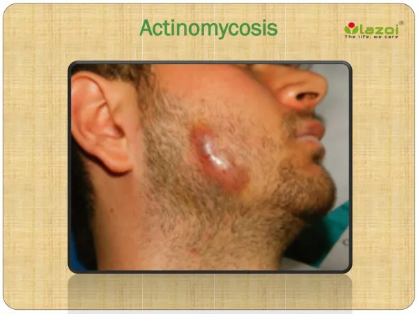
Disease: actinomycosis • the most common -- Actinomyces israelii • endogenous organism • tooth abscess or suppurative infections in thoracic area and abdomen • a pus-draining lesion
diagnosis -- "sulfur granules" • The yellow granules can be seen with the naked eye.
diagnosis -- "sulfur granules" • Microscopy: G+ bacillus with branching filaments, sulfur granules are surrounded by polymorphonuclear cells (PMN), forming the purulent tissue reaction.
treatment • These lesions must be surgically drained prior to antibiotic therapy and the drug of choice is large doses of penicillin (2 million units q 6 h).
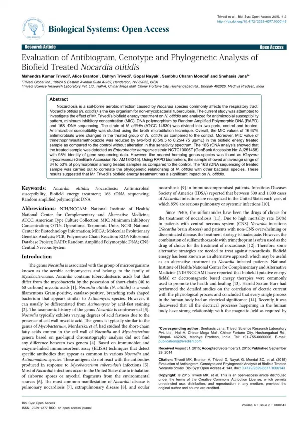
Nocardiosis • Subcutaneous infections, pulmonary infections, and brain abscesses • N. asteroides; N. brasiliensis
Lab. Diagnosis • Clinical Specimens: sputum, pus, biopsy tissue
Treatment Sulfamethoxazole-Trimethoprim
Case study I • A 58-year old lawyer presents in the emergency room with headache, irritability, generalized muscle pain and uncontrollable back spasms. He has become very restless and worried because he has had the back spasms all through his court case that afternoon and they became extremely painful.
In his history, the lawyer states that he has a very busy practice. He is on medication for high blood pressure (beta blocker) and has mild asthma. He injured himself about 10 days earlier, puncturing his left arm with a nail from an old barn he is tearing. The wound has produced moderate quantities of pus, but he has been keeping it clean. When asked, he did not remember having a tetanus shot since he was a kid. • The wound was sampled for microscopic examination and culture. The back appears to have very tight contractions and spasms. The patient is in obvious agonizing pain. • What is your diagnosis here? • What is the proper treatment of this problem? • How could this have been prevented?
Case study II • A 62-year old diabetic black man presents in the emergency room with a swollen left leg with areas of blanching and blue mottling. A "foul odor" is coming from a dressed wound. The physicians remove the dressing and a brownish fluid is seeping from a wounded area. The fluid contains what appear to be small bits of the tissue. No pus appears to be present. The wound has a strong "rotten" odor.
Five days earlier, while at his work as a farmer, he caught the leg in his manure spreader, sustaining a deep, crushing, grossly dirty injury. His wife cleaned the wound as well as she could with soap and water, dressed it with clean gauze, and wrapped it tightly with an elastic bandage to stop the bleeding. The second day they redressed the wound and applied triple antibiotic ointment. The patient treated his pain with ibuprofen (Advil). He reported the pain was not very bad for the first 72 hours. In the past 24 hours, the leg swelled and the mottling began to appear. A foul odor and severe pain accompanied the swelling. His wife convinced him to come to the emergency room even though they did not have medical insurance.
What is your diagnosis in this case? • How should this wound be treated? • Is this a life-threatening condition? • Is it likely that the patient’s diabetes contributed to the problem as presented?
Case study III • A 68-year old patient with Alzheimer disease was brought to the emergency room by the staff of a local nursing home. He presented as lethargic with a sallow complexion. He had an admission temperature of 102.4F and a respiratory rate of 33/minute. During respiration, the right side of his chest moved better than the left. He showed dense consolidation of the lower lobe of the left lung on physical exam. A sputum sample revealed blood and a greenish color.
A chest x-ray showed tight consolidation of the left lung with evidence of formation of cavities in the lung tissue from cytotoxic damage. The patient complained of chills in the exam room, combined with his fever. A smear of his sputum demonstrated no acid-fast bacteria. • What is your presumptive diagnosis for this case? • What evidence could the sputum give for this case? • Is the reduced respiration rate and unequal chest movement indicative of the pathology? • Is this a bacterial or viral disease?