Download
1 / 42
540 likes | 1.24k Vues
db -Thalassemia. Disorders of Hemoglobin Dr. Pupak Derakhshandeh. Structure and function of hemoglobin. Oxygen carrier In vertebrate: red blood cells Four subunits: 2 α - and 2 -chains. Understanding globin regulation in β-thalassemia: it’s as simple as α, β, γ, δ. Arthur Bank

E N D
db-Thalassemia Disorders of Hemoglobin Dr. Pupak Derakhshandeh
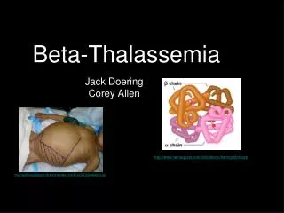
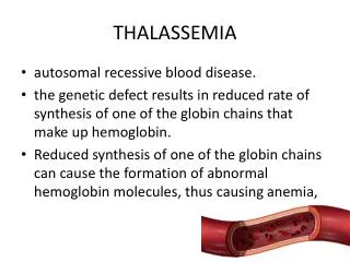
Structure and function of hemoglobin • Oxygen carrier • In vertebrate: red blood cells • Four subunits: • 2α- and 2-chains
Understanding globin regulation in β-thalassemia:it’s as simple as α, β, γ, δ Arthur Bank The Journal of Clinical Investigation http://www.jci.org Volume 115 Number 6 June 2005
The human globin loci • The best characterized in the human genome at the gene and protein levels • The β–locus control region (β-LCR): • A dominant control region located upstream of the globin structural genes • a strong enhancer of the expression of the downstream
.The human globin locus and their role in β-thalassemia(A) The β-LCR and structural genes (ε, Gγ, Aγ, δ, and β) in the β-globin locus on chromosome 11
The major genes expressed throughout fetal life • The α-globin gene • 2 γ-globin genes, Gγ and Aγ
(B) The α-globin locus is shown with the ζ- and 2 α-globin genes on chromosome 16
The δ- and β-globin • activated late in fetal life • with the β-globin gene • most highly expressed in erythroid cells during adult life
b-Globin gene expression • between cis-acting sequences: • The β-LCR • trans-acting factors: • including transcription factors
Tetramers of globin chains • The most stable configuration of hemoglobin • associated with heme groups
(C) In early fetal life, the α- and γ-globin chains combine to form HbF (α2γ2), the main β-globin–like globin during the remainder of fetal life and early postnatal life Severe anemia results >>
The current therapy for β-thalassemia • Blood transfusions + iron Chelation • Decreasing α-globin accumulation • and/or reactivating γ-globin production • BM transplantation
Decreasing excess α-globinaccumulation • Unequal crossing over in meiosis: • deletion of the α-globin gene • reduces α-globin synthesis in patients • Homozygous for β-thalassemia (Major) + decreases the α-globin excess >> • decreased severity of anemia
Increasing human γ-globinexpression • reduce anemia and cure human β-thalassemia • increase in human γ-globin gene expression >> restoration of HbF • Point mutations in the γ-globin gene promoter: • increase γ-globin expression, but not by agreat amount
Hereditary persistence of fetal hemoglobin (HPFH) • express γ-globin genes at the same level in adult life as in fetal life • Some HPFH homozygotes have only HbF (a2g2) and no anemia!
Doesn't cause any health problem • HPFH / Thalassemia (no problem) • HPFH / HPFH
HPFH as a δβ-globinDisease • Large deletions at the β-globin locus • from the region close to the human Aγ gene to well downstream of the human β-globin • gene and including deletion of the structural δ- and β-globin genes
HPFH • Heterozygotes: • a normal level of HbA2 • even higher levels of HbF (15 to 30 %) • Homozygotes: • clinically normal • albeit with reduced MCV and MCH • Compound heterozygotes with b thalassemia: • clinically very mild
HPFH • group of disorders • characterized by a decreased or absent: • b-chain synthesis • a variable compensatory increase in g-chain synthesis
Intergenic γδ sequences: γ-globin gene regulation • Corfu: • homozygous for the Corfu deletion • a deletion of 7.2 kb DNA • upstream of the δ-globin • homozygotes were shown to possess 88%-90% HbF • only mild anemia • Did not require blood transfusion
A guide to the diagnosis of the different forms of haemoglobinopathies in carriers (Cao et al., 2001)
Molecular diagnosis of haemoglobin disordersClin. Lab. Haem. 2004, 26, 159–176B. E. CLARK, S. L. THEINDepartment of Haematological Medicine, King’s College Hospital and GKT School of Medicine,Denmark Hill, London, UK
The beta locus on chromosome 11 p15.4 with the e,Gg and Ag, d and b genes, arranged in the order of their developmental expression
Gap-PCR db-thalassaemias: • the common HPFH • Hb Lepore -a Thalassemia, …
Gap-PCR for the African HPFH-2 deletion N D N N D N N D D D N D D N N 918 639 bp
Multiplex gap-PCR for detection of the common a-thalassaemia deletions (-/a3.7/-a4.2) (- -SEA/aa) (aa/aa) (a20.5/a3.7) (- -MED/a3.7)
Homozygosity for nondeletion db0 thalassemia resulting in a silentclinical phenotype BLOOD, 1 SEPTEMBER 2002 VOLUME 100, NUMBER 5 Renzo Galanello, Susanna Barella, Stefania Satta, Liliana Maccioni, Carlo Pintor, and Antonio Cao
Nondeletion Sardinian db0 thalassemia • a homozygous state for nondeletion Sardinian db0 thalassemia • a symptomless clinical phenotype with • pattern (Hb F: 99.8% and Hb A2: 0.2%)
The molecular defects • the presence of 2 nucleotide substitutions: • -196C>T in the promoter of the Ag-globin gene • 39C>T nonsense mutation in b-globin gene * *
The absence of typical thalassemia clinical findings • high Hb F output: which compensated for the absence of chains • The near absence of Hb A2: • alterations in the globin gene transcriptional : • Activation of g-globin genes • and suppression of d-globin genes • or preferential survival of red blood cells with the highest Hb F • and low Hb A2 level
The absence of typical thalassemia clinical findings • The imbalance in the ratio of a to g • similar to that in heterozygous thalassemia • explains the reduction in MCV • mean corpuscular Hb
Patient with nondeletion homozygous db0 thalassemia • Had almost no HbA2 (0.3%) • the suppressive effect of the in cis Ag -196CT mutation • This suppressive in cis effect has already been reported for similar mutations, such as the -202 Gg HPFH