Download
1 / 22
300 likes | 990 Vues
SKIN, HAIR, NAIL AND MAMMARY GLAND. Dr. Andrea D. Székely. SKIN APPENDAGES. HAIR NAILS GLANDS. I. II. III. SKIN - INTEGEMENTUM COMMUNE. epidermis. I. epidermis II. dermis, corium III. tela subcutanea, subcutis, hypodermis. str. corneum. str. lucidum (eleidin). str. granulosum.

E N D
SKIN, HAIR, NAIL AND MAMMARY GLAND Dr. Andrea D. Székely
SKIN APPENDAGES HAIR NAILS GLANDS
I. II. III. SKIN - INTEGEMENTUM COMMUNE epidermis I. epidermis II. dermis, corium III. tela subcutanea, subcutis, hypodermis str. corneum str. lucidum (eleidin) str. granulosum (keratohialin) str. spinosum str. germinativum (basale) Sweat gland, excretory duct • - Keratinocyte • * produces keratin • * 27-30 days cycle • - Melanocyte • * produces melanin from tyrosin (tyrosinase enzyme) • - Merkel cell • * binds the touch receptor to a Nerve • Langerhans cell • * Antigene presentation str. lucidum str. granulosum str. spinosum Keratinocytes Langerhans cell str. basale melanocyte Merkel cells
plexus subpapillaris I. a II. b III. rete corii INTEGEMENTUM COMMUNE I. epidermis II. dermis, corium III. tela subcutanea, subcutis, hypodermis DERMIS, CORIUM) a.: stratum papillare b.: stratum reticulare • - Fine fibrous structure (coll+elast) • - plexus venosus subpapillaris • - CT papillae against the epidermis • - number of papillae - support • the Epithelium follows the Papillae • * cristae cutis • * sulci cutis • * toruli tactiles (Finger tip) • - Strong collagen fibres + • elastic network • - Hair follicles • - glands, vessels • - CT cells • Mobile Elements of the • Immune system • - Nerves, Receptors TELA SUBCUTANEA, SUBCUTIS, HYPODERMIS • - Connection between skin and CT • Gives the skin a certain mobility • Stress tolerance • - Difference in thickness • - Rich in fat lots of CT fibres (retinacula cutis) • (panniculus adiposus): Fat depo; Isolator
I. II. III. Ruffini Krause Meissner INTEGEMENTUM COMMUNE AS A SENSORY ORGAN 1. free nerve endings 2. follicular afferets 3. Skin receptors - Merkel’s touch corpuscle - Meissner’s – ” - - Vater-Paccini – „ - (stretch and vibration) - Ruffini’s corpuscle (stretch, temperature) - Krause’s corpuscle (stretch, cold receptor) 4. Vegetative fibres: sudo-, vaso-, pilomotor axons Freie Nervendigungen Vater-Paccini Merkel
I. II. III. INTEGEMENTUM COMMUNE – GLANDS 1. (GLANDULAE CUTIS) Merocrine secretion SWEAT GLANDS (glandulae sudoriferae) true, eccrine type SECRETION eccrine Sweat glands apocrine GLANDS AXILLA apocrine secretion
I. II. III. INTEGEMENTUM COMMUNE –GLANDS 2. (GLANDULAE CUTIS) 1. SALIVARY GLANDS (GLANDULAE SUDORIFERAE) - TRUE, ECCRINE TYPE -APOCRINE TYPE SWEAT GLANDS (GLL ODORIFERAE) 2. SEBACEOUS GLAND (GLANDULAE SEBACEAE) A 2 B 1 HOLOCRINE SECRETION 2. SEBACEOUS GLANDS (GLANDULAE SEBACEAE)
Babies haven't any hair;Old men's heads are just as bare;Between the cradle and the graveLies a haircut and a shave. --Samuel Hoffenstein (1890-1947)Songs of Faith in the Year After Next HAIR 1. • DISTRIBUTION • ALL OVER, except : the palms, the soles, the lips • Hair is most obvious on the head and face (including the nose and ears in some people), • the armpits, the groin, and (in men) the chest and legs. • How much hair do we have? • On average, a head carries about 100000 hair follicles. Some people have as many as 150,000. • baby's head cca 1100 follicles/ cm2. • By the age of 25, this number is reduced to 600, (depends on the physical type) • Between the ages of 30 and 50, it is further reduced to 250-300. (and so it continues…) • Each follicle grows about 20 new hairs in a lifetime. Each new hair grows for several years, and can reach over a metre in length. • Each hair falls out eventually, and is replaced by a new one TYPES terminal lanugo • vellus
HAIR2. COMPOSITION keratin (dead keratinocytes), fat, pigment (melanin), small amounts of vitamins, traces of zinc or other metals, water (10-13%) PARTS shaft (above the skin) root (below the surface). follicle (indentation of the epidermis) sebaceous gland arrector pili (WHY: keep warm or look bigger to impress the other sex or intimidate enemies) COLOUR due to melanin, (melanocytes in the bulb of the hair follicle) - Dark hair - contains true melanin, blond and red hair result from types of melanin that contain sulfur and iron - Gray hair - melanocytes age and lose the enzyme necessary to produce melanin. - White hair occurs when air bubbles become incorporated into the growing hair. TEXTURE (defined by the shape of the hair shaft) straight hair - round in cross section, wavy hair - oval shape in cross section, curly hair - elliptical or kidney-shaped
HAIR FOLLICLE – HISTOLOGY Connective tissue Connective tissue external Connective tissue internal external Cu external M internal Cu Cortex Cortex M M
HAIR FOLLICLE – EMBRYOLOGY The dermal papilla (DP)induces the hair follicle and retains this instructive ability throughout the life of a hair follicle (removal of the DP stops hair growth but the lower third of the dermal sheath induces the regeneration of a new DP (hair follicle regrowth). The DP cells retain their embryonic functional abilities and are able to induce new hair fibre growth in mature, adult skin when implanted into previously deactivated hair follicles and in close association with ORS epidermal cells.
HAIR GROWTH Hair growth occurs in a cycle. Phases of the hair growth cycle; anagen, catagen and telogen with anagen further subdivided into proanagen, mesanagen and metanagen. Anagen is the active growth phase when hair fibre is produced. Proanagen marks initiation of growth with RNA and DNA synthesis in a follicle which then quickly progresses through mesanagen to metanagen and maximum follicle length and girth. In this mature state of proliferation and differentiation the hair follicle consists of a total of eight concentric layers and melanogenesis occurs within pigmented hair follicles. Anagen is followed by catagen, a period of controlled regression of the hair follicle. Ultimately the hair follicle enters telogen, when the follicle is in a so-called resting state.
NAIL – GENERAL OVERVIEW Nails are flattened, elastic structures of a horny texture that protect the tips of the terminal phalanges (toes and fingers). Convex on the outer surface and concave within, human nails are implanted by their root into a groove in the skin (nail sulcus). The nail matrix, underlying the body and root of the nail, is the source of new nail production. The white part of the nail, the lunula, represents the portion of the nail that is not firmly attached to the connective tissue base and contrasts with the redder, highly vascularized majority of the nail that is attached to the thick matrix. Cuticles are continuous with the horny substance of the nail as part of the epidermis. Nails grow in length by producing new cells at the root of the nail, and at the distal free edge, the oldest nail cells reside.
margo liber hyponychium Nail plate Nail bed (sinus unguis) lunula eponychium phalanx distalis proximal nail fold matrix unguis margo liber Nail plate Nail bed (sinus unguis) eponychium lateral nail groove Lateral Nail fold corpus unguis lateral Nail fold lunula phalanx distalis Fibres to Dermis Nail plate Below the proximal Nail fold (radix unguis) eponychium NAIL (UNGUIS, ONYX)
NAIL – HISTOLOGY AND EMBRYOLOGY nail fold (NF), the matrix region of the nail root (M), the nail bed (NB), nail proper (N), eponychium (Ep) and hyponychium (Hy), cartilage (C), epiphysial plate (EpPl), bone (B) week 10 of human development, the fetus develops its fingernails week 14 the ten toenailsfollow Originally, the nail fields appear at the tips of the digits and then migrate toward the dorsal surfaces. While the surrounding cells form the nail folds, keratinization of the proximal nail folds forms the nail plates. By week 32, the fingernails, and by week 36, toenails, reach the tips of the digits. (indicator of the degree of maturity or prematurity)
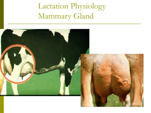
Anterior axillary fold m. pectoralis major Anterior axillary fold; m. pectoralis major costa IV papilla mammae areola mammae costa VI papilla mammae areola mammae glandulae areolares; Montgomery glands (5-15 St) m. serratus anterior mamma MAMMA
fascia pectoralis spf. lobuli glandulae mammae m. pectoralis major corpus adiposum mammae m. pectoralis major fascia pectoralis prof. cutis mammae Vordere Achselfalte m. pectoralis major retinacula cutis mm. intercostales sinus lactiferi glandula mammae costa IV papilla mammae lobus mammae 14-24 db mamilla costa VI m. serratus anterior areola mammae porus lactiferus lobulus mammae corpus mammae; glandula mammae sinus lactiferus areola mammae: glandulae areolares m. arrector mammae ductus lactiferus m. serratus anterior papilla mammae: porus lactiferus (15-20) MAMMA • paired organs • regional borders: sternum and anterior axillary fold • in males - reduced (mamma virilis) – HOWEVER – breast cancer is possible!!! • consists of glandular lobes (glandula mammae) and dense fatty CT (corpus adiposum mammae) • produces milk
MAMMA ARTERIES: - a. thoracica interna - a. thoracica lateralis - aa. intercostales posteriores VEINS: - v. thoracica interna - v. thoracica lateralis - vv. intercoastales posteriores - v. thoracoepigastrica NERVES: - nn. supraclaviculares - nn. intercostales
nullipara pregnancy Mammalactans MAMMA Mamma non lactans Mamma lactans Apocrine secretion
MAMMA Ultraultrasoundschall Mammography nodi lymphatici deltoideopectorales (infraclaviculares) nodi lymphatici axillares nodi lymphatici pectorales nodi lymphatici parasternales Self examination
PLASTIC SURGERY OF THE BREASTS BREAST REDUCTION FOLLOWING BREAST REMOVAL - RECONSTRUCTING SURGERY SLIPPED IMPLANT