PROVIDED BY EMAD BEHDAD
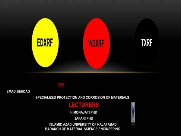
EDXRF. WDXRF. TXRF. PROVIDED BY EMAD BEHDAD SPECIALIZED PROTECTION AND CORROSION OF MATERIALS LECTURERS H.MONAJATI,PHD JAFARI,PHD

PROVIDED BY EMAD BEHDAD
E N D
Presentation Transcript
EDXRF WDXRF TXRF PROVIDED BY EMAD BEHDAD SPECIALIZED PROTECTION AND CORROSION OF MATERIALS LECTURERS H.MONAJATI,PHD JAFARI,PHD ISLAMIC AZAD UNIVERSITY OF NAJAFABAD BARANCH OF MATERIAL SCIENCE ENGINEERING
INTRODUCTION X-ray Fluorescence (XRF) • Atoms in the sample are excited and emit characteristic x-rays x-ray source x-ray detector Types of elements present (qualitative analysis) Number of x-rays for each element concentration (quantitative analysis)
Types of XRF instruments Based on the excitation • • Tube excited XRF • • Radio-isotope excited XRF • • Secondary target • • Synchrotron, • • Total reflectionof x-ray florescence • • Wavelength dispersive (WD-XRF) • • Energy dispersive (ED-XRF) • • Filter instruments (proportional counter) Based on the detection
What is edx ? • *Energy dispersive X-ray spectroscopy (EDS or EDX) • *Analytical technique used for the elemental analysis • *Technique used for chemical characterization of a sample • *Investigation of a sample • *Analyzing X-rays emitted by the matter • *Full quantitative analysis showing the sample composition
How edx works ? • A high-energy beam of charged particles is focused into the sample • Ground state(unexcited) electrons in sample are stimulated • Electrons are excited from lower energy shells to higher energy shell • The difference in energy between the shells may be released in the form of an X-ray • The number and energy of the X-rays emitted from a specimen can be measured by an energy dispersive spectrometer
Energy-dispersive spectrometers What is the principle behind ED spectrometers? The heart of an ED-spectrometer is a semi-conductor crystal (Si, Ge) a high voltage is applied over the crystal (bias -600V) and the crystal is cooled (e.g. at liquid nitrogen temperature) When x-rays enter the crystal electron-hole pairs are formed the number is proportional to the energy of the x-ray because of the bias the electrons are swept out of the crystal For each photon an electric pulse is produced with an amplitude proportional to the energy. Measuring the amplitude and counting produces the ED-spectrum.
WHY edx • Fast • (Non)-destructive • Sample Area • Good precision, accuracy • Fair sensitivity • Multi-element analysis • Generally not portable • Some sample preparation required • Experienced staff • Moderately expensive
What is wdx ? • Wavelength dispersive X-ray spectroscopy (WDXRF or WDS) • A method used to count the number of X-rays • Reads or counts only the x-rays of a single wavelength • Element must be known • Often used in conjunction with EDS
Usage areas of wdx • Identification of spectrally overlapped elements • Detection of low concentration species (10-100 ppm) • Analysis of low atomic number elements • Oxidation and corrosion of metals
How wdx works • The WDX operates in much the same way as EDX. • Unlike the related technique of Energy dispersive X-ray spectroscopy (EDX)WDX reads or counts only the x-rays of a single wavelength, not producing a broad spectrum of wavelengths or energies. • The crystal structure of sample diffracts the photons in principles of Bragg's law. • Diffractions are then collected by a detector.
WHY WDX • (Non)-destructive • High accuracy • Excellent precision & long term stability • Good resolution & sensitivity • Multi-element analysis • Well-established technique • Some sample preparation • Qualified, experience staff • Expensive
Comparison of WD and ED X-ray Detectors • Most important advantages of WD: Higher resolution, sensitivity • Most important advantages of ED: Cheaper, faster (except for multichannel WD) • Other differences (more detailed comparison):
Total Reflection X-Ray Fluorescence (TXRF) • Total reflection X-ray fluorescence analysis (TXRF) is basically an energy dispersive • analytical technique in special excitation geometry.
Usage areas of txrf • Total reflection X-ray fluorescence (TXRF) has become increasingly popular in micro and trace elemental analysis. • It is being used in geology, biology, materials science, medicine, forensics, archaeology, art history, and more. • Unlike the high incident angles (~ 40 °) used in traditional XRF, TXRF involves very low incident angles. • These low angles allow the X-rays to undergo total reflection.
Advantages of TXRF • • Background reduced. • • Double excitation of sample by both the primary and the reflected beam. • • No matrix effects. • • Calibration and quantification independent from any sample matrix. • • Simultaneous multi-element ultra-trace analysis. • • Several different sample types and applications. • • Excellent detection limits (ppt or pg) for all elements from sodium to plutonium. • • Excellent dynamic range from ppt to percent. • • Possibility to analyses the sample directly without chemical pretreatment. • • Non destructive analysis. • • Low running cost.
Experimental analysis results Spectrum of a 3 μL mineral water sample, spiked with 1 ng/μL Ga as internal standard element. Excitation in TXRF geometry with a multilayer monochromator by a Mo X-ray tube (50 kV, 10 mA, 1000 s measuring time).
PIXEPROVIDED BY ZABIH ALLAH KHANSHA Proton Induced X-ray Emission
Interaction between MeV ions and a material surface.Processes relevant to ion beam analysis
Proton Induced X-ray Emission Detector Si(Li) X-rays 2-3 MeV sample
Pixe usage 2004 16% 33% 27% 24%
Typical PIXE spectrum of normal and varicose veins with 175mm Maylar absorber in front of Si(Li) detector
QUESTIONS ? • THANKS FOR YOUR ATTENTION