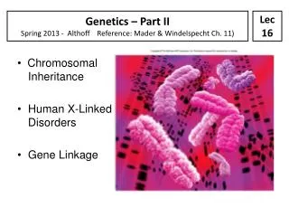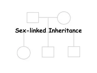
X-Linked Inheritance
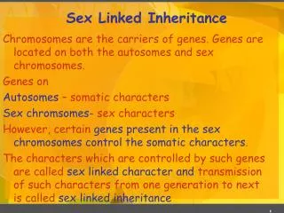
Lecture 7. X-Linked Inheritance. X-linked recessive disorders Responsible gene on X chromosome For females, both copies of the X chromosome must be affected Males, hemizygous for the X chromosome, much more likely to be affected. Some Common Sex-Linked Recessive Disorders.

X-Linked Inheritance
E N D
Presentation Transcript
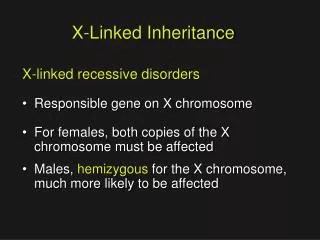
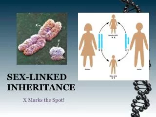
Lecture 7 X-Linked Inheritance X-linked recessive disorders • Responsible gene on X chromosome • For females, both copies of the X chromosome must be affected • Males, hemizygous for the X chromosome, much more likely to be affected
Some CommonSex-Linked Recessive Disorders • Duchenne and Becker Muscular Dystrophy • Hemophilia A • Glucose-6-phosphate dehydrogenase deficiency • Fragile X Mental Retardation • Color blindness
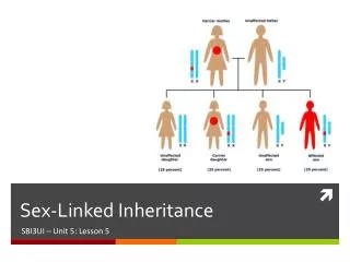
Maternal X x XX Xx X 1/4 1/4 Paternal XY Y XY 1/4 1/4 F M unaffected non-carrier 1/2 1/2 unaffected carrier 1/2 affected 1/2 Hemophilia A Predict possible fetal outcomes
Haemophilia AAn X-linked recessive disease • Caused by mutation in the clotting factor VIII gene (F8) on chromosome Xq28 • Incidence: 1/5,000 males births
Clinical symptoms Haemorrhage into joints and muscles, easy bruising, and prolonged bleeding from wounds.
X-inactivation, Dosage compensation, and the expression of X-linked genes • Males • One X chromosome • Females • Two X chromosomes • And yet, the mean amounts of gene products of X-linked genes are the same in males as in females • HOW? • Through X chromosome inactivation Same amount of X-linked gene products between males and females
The molecular mechanism behind X-inactivation • The key player is the X-linked gene XIST • X(inactive)-specific transcript • Chromosome Xq13.2 • XIST is transcribed to produce a non-coding RNA that “coats” the X-chromosome and inactivates it • XIST is uniquely expressed from the inactive X • XIST RNA does not travel over to any other X chromosome in the nucleus. • Barr bodies are inactive X chromosomes "painted" with XIST RNA.
Transcription of XISTceases on the other X chromosome allowing all of its hundreds of other genes to be expressed. The shut-down of the XIST locus on the active X chromosome is done by methylating XIST regulatory sequences. • So methylation permanently blocks XIST expression and permits the continued expression of all the other X-linked genes.
The XIST gene on one of the two X-chromosomes is randomly inactivated by DNA methylation The active XIST is transcribed and its RNA product coats the X-chromosome XIST The histones on the coated X undergo methylation which causes the chromosome to condense, forming a Barr body, and renders it inactive X with inactive XIST X with active XIST The uncoated X is left transcriptionally active Barr body
Expression of X-linked Genes in Heterozyotes • Inactivation is random, established when embryo < 100 cells fraction of cells in carrier female with normal or mutant allele tend to be variable • Thus, clinical variation in expression of X-linked disorders is common in heterozygotes ranging from normal to affected • A manifesting heterozygote is a female in whom the deleterious allele is on the active X in most or all of cells (an extreme e.g., of unbalanced or skewed X-inactivation)
X chromosome Inactivation • Inactivation is not always random • A structurally abnormal X is preferentially inactivated, e.g., isochromosome X • E.g., extraembryonic membranes (that go on to form the amnion, placenta, and umbilical cord). In all the cells of the extraembryonic membranes, it is father's X chromosome that is inactivated. • Inactivation is not complete • Some genes are known to escape inactivation (i.e. those with a functional homolog on the Y, e.g., genes located in the pseudoautosomal region) • Inactivation is not permanent • reversed in development of germ cells (not passed on to gametes)
X-autosome translocation There is normally a 50% chance that a particular X will be inactivated in a cell from a female If an X bears a piece of autosome(translocation) then the untranslocated X is always inactivated since the cell needs both copies of the autosomal genes to be active If the translocated X has a mutant allele, all the woman’s cells will be mutant
Functional Mosaicism Resulting from X-inactivation • Females are mosaics wrt their X-linked genes • Mosaicism is readily detected for some disorders e.g., DMD
Immunostaining for dystrophin in muscle specimens. A, A normal female (magnification ×480). B, A male with Duchenne muscular dystrophy (×480). C, A carrier female (×240). Staining creates the bright lines seen here encircling individual muscle fibers. Muscle from DMD patients lacks dystrophin staining. Muscle from DMD carriers exhibits both positive and negative patches of dystrophin immunostaining, reflecting X inactivation
Example: hemophilia A Predict possible fetal outcomes P
Homozygous Affected Females Consanguinity in an X-linked recessive pedigree for red-green color blindness, resulting in a homozygous affected female
New Mutation in X-linked Disorders • For a sex-linked recessive disorder with zero fitness, such as Duchenne muscular dystrophy, 1/3 of disease alleles are in males and are lost with each generation. Thus, 1/3 of disease alleles must be replaced with a new mutation in each generation • DMD is said to be genetic lethal because affected males usually fail to reproduce • For hemophilia, in which reproduction is reduced but not eliminated, a proportionately smaller fraction of cases will be due to new mutation
Characteristics of Sex-LinkedRecessive Inheritance • Males are more commonly affected than females. • The gene responsible is transmitted from an affected man through his daughters, who are seldom affected. Each daughter is an obligatory heterozygous carrier. Each of the carrier daughter's sons has a 50% chance of inheriting it. • No male to male transmission occurs. • The affected males in a pedigree are usually related through females. • Heterozygous female carriers are usually unaffected, but some may express the condition with variable severity (“Lyonization”).
X-Linked Dominant Inheritance • Responsible gene on X chromosome • The phenotype is regularly expressed in heterozygotes • Affected fathers transmit the disorder to ALL of their daughters none of their sons • The pattern of inheritance through females is no different from AD pattern • Each child of an affected female has a 50% chance of inheriting the trait, regardless of sex • Rare X-linked dominant phenotypes are about twice as common in females, though the expression is much milder in females who are almost always heterozygous
X-Linked Dominant Inheritance • X-linked hypophosphatemic rickets, also called vitamin D-resistant rickets, in which ability of kidney tubules to reabsorb filtered phosphate is impaired • Serum phosphate level is less depressed and rickets less severe in heterozygous females as compared to affected males • The defective gene product appears to be a member of a family of endopeptidases, but the pathogenic mechanism is not known
X-linked Dominant Disorders with Male Lethality • Some rare genetic defects expressed exclusively or almost exclusively in females appear to be XD lethal in males before birth • Typical pedigrees: transmission by affected female affected daughters, normal daughters, normal sons in equal proportions (1:1:1) • Rett syndrome meets criteria for an XD that is usually lethal in hemizygous males. The syndrome is characterized by normal prenatal and neonatal growth and development, followed by rapid onset of neurological symptoms and loss of milestones between 6 and 18 months of age.
Rett syndrome cont. • Children become spastic and ataxic, develop autistic features and irritable behavior with outbursts of crying, and demonstrate characteristic purposeless wringing or flapping movements of hands and arms. • Head growth slows and microcephaly develops. Seizures are common (~50%) • Mental deterioration stops after a few years and the patients can then survive for many decades with a stable but severe neurological disability. • Most cases caused by spontaneous mutations in an X-linked MECP2 gene encoding methyl CpG binding protein 2. ? Thought to reflect abnormalities in regulation of genes in developing brain.
Typical appearance and hand posture of girls with Rett syndrome
Rett syndrome cont. • Males who survive with the syndrome usually have two X chromosomes (as in 47,XXY or in a 46,X,der(X) male with the male determining SRY gene translocated to an X) or are mosaic for a mutation that is absent in most of their cells • There are a few apparently unaffected women who have given birth to more than one child with Rett syndrome. ? X-inactivation pattern in a heterozygous female. ? Germline mosaic
Pedigree pattern demonstrating an X-linked dominant disorder, lethal in males during the prenatal period.
Characteristics of X-Linked Dominant Inheritance • Affected fathers with normal mates have no affected sons and no normal daughters • For rare pehnotypes, affected females are about twice as common as affected males (unless disease is lethal in males), but affected females typically have milder (though variable) expression • Both male and female offspring of a heterozygous female have a 50% risk of inheriting the phenotype. The pedigree pattern is similar to AD inheritance
Patterns of Pseudoautosomal Inheritance • Genes on pseudoautosomal region can regularly exchange b/w the two sex chr’s • E.g., Dyschondrosteosis, a dominantly inherited skeletal dysplasia with disproportionate short stature and deformity of the forearm • The responsible gene is pseudoautosomal that escapes X-inactivation, encodes a transcription factor likely involved in stature • Either deletion/mutations dyschondrosteosis in both heterozygous males and females
Inheritance pattern of dyschondrosteosis. Arrow shows a male who inherited the trait on his Y chr. from his father. His father, however, had inherited the trait on his X chr. from his mother