The Muscular System
350 likes | 425 Vues
Delve into the complexities of the muscular system, exploring different muscle types, their functions, and unique characteristics. From skeletal to cardiac and smooth muscles, discover how muscles work and contribute to bodily movement and stability.

The Muscular System
E N D
Presentation Transcript
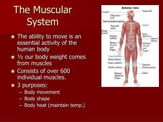
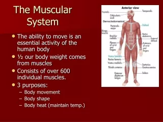
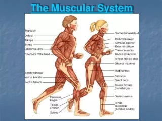
The Muscular System • Muscles are responsible for all types of body movement • Three basic muscle types are found in the body • Skeletal muscle • Cardiac muscle • Smooth muscle
Characteristics of Muscles • Skeletal and smooth muscle cells are elongated (muscle cell = muscle fiber) • Contraction of muscles is due to the movement of microfilaments • All muscles share some terminology • Prefixes myo and mys refer to “muscle” • Prefix sarco refers to “flesh”
Skeletal Muscle Characteristics • Most are attached by tendons to bones • Cells are multinucleate • Striated—have visible banding • Voluntary—subject to conscious control
Connective Tissue Wrappings of Skeletal Muscle • Cells are surrounded and bundled by connective tissue • Endomysium—encloses a single muscle fiber • Perimysium—wraps around a fascicle (bundle) of muscle fibers • Epimysium—covers the entire skeletal muscle • Fascia—on the outside of the epimysium
Skeletal Muscle Attachments • Epimysium blends into a connective tissue attachment • Tendons—cord-like structures • Mostly collagen fibers • Often cross a joint due to toughness and small size • Aponeuroses—sheet-like structures • Attach muscles indirectly to bones, cartilages, or connective tissue coverings
Skeletal Muscle Attachments • Sites of muscle attachment • Bones • Cartilages • Connective tissue coverings
Smooth Muscle Characteristics • Lacks striations • Spindle-shaped cells • Single nucleus • Involuntary—no conscious control • Found mainly in the walls of hollow organs
Smooth Muscle Characteristics Figure 6.2a
Cardiac Muscle Characteristics • Striations • Usually has a single nucleus • Branching cells • Joined to another muscle cell at an intercalated disc • Involuntary • Found only in the heart
Cardiac Muscle Characteristics Figure 6.2b
Skeletal Muscle Functions • Produce movement • Maintain posture • Stabilize joints • Generate heat
Microscopic Anatomy of Skeletal Muscle • Sarcolemma—specialized plasma membrane • Myofibrils—long organelles inside muscle cell • Sarcoplasmic reticulum—specialized smooth endoplasmic reticulum
Microscopic Anatomy of Skeletal Muscle Figure 6.3a
Microscopic Anatomy of Skeletal Muscle • Myofibrils are aligned to give distinct bands • I band = light band • Contains only thin filaments • A band = dark band • Contains the entire length of the thick filaments
Microscopic Anatomy of Skeletal Muscle Figure 6.3b
Microscopic Anatomy of Skeletal Muscle • Sarcomere—contractile unit of a muscle fiber • Organization of the sarcomere • Myofilaments • Thick filaments = myosin filaments • Thin filaments = actin filaments
Microscopic Anatomy of Skeletal Muscle • Thick filaments = myosin filaments • Composed of the protein myosin • Has ATPase enzymes • Myosin filaments have heads (extensions, or cross bridges) • Myosin and actin overlap somewhat • Thin filaments = actin filaments • Composed of the protein actin • Anchored to the Z disc
Microscopic Anatomy of Skeletal Muscle Figure 6.3c
Microscopic Anatomy of Skeletal Muscle • At rest, there is a bare zone that lacks actin filaments called the H zone • Sarcoplasmic reticulum (SR) • Stores and releases calcium • Surrounds the myofibril
Microscopic Anatomy of Skeletal Muscle Figure 6.3d
Stimulation and Contraction of Single Skeletal Muscle Cells • Excitability (also called responsiveness or irritability)—ability to receive and respond to a stimulus • Contractility—ability to shorten when an adequate stimulus is received • Extensibility—ability of muscle cells to be stretched • Elasticity—ability to recoil and resume resting length after stretching
The Nerve Stimulus and Action Potential • Skeletal muscles must be stimulated by a motor neuron (nerve cell) to contract • Motor unit—one motor neuron and all the skeletal muscle cells stimulated by that neuron
The Nerve Stimulus and Action Potential Figure 6.4a
The Nerve Stimulus and Action Potential Figure 6.4b
The Nerve Stimulus and Action Potential • Neuromuscular junction • Association site of axon terminal of the motor neuron and muscle
The Nerve Stimulus and Action Potential Figure 6.5a
The Nerve Stimulus and Action Potential • Synaptic cleft • Gap between nerve and muscle • Nerve and muscle do not make contact • Area between nerve and muscle is filled with interstitial fluid
Transmission of Nerve Impulse to Muscle • Neurotransmitter—chemical released by nerve upon arrival of nerve impulse • The neurotransmitter for skeletal muscle is acetylcholine (ACh) • Acetylcholine attaches to receptors on the sarcolemma • Sarcolemma becomes permeable to sodium (Na+)
Transmission of Nerve Impulse to Muscle • Sodium rushes into the cell generating an action potential • Once started, muscle contraction cannot be stopped
Transmission of Nerve Impulse to Muscle Figure 6.6
The Sliding Filament Theory of Muscle Contraction • Activation by nerve causes myosin heads (cross bridges) to attach to binding sites on the thin filament • Myosin heads then bind to the next site of the thin filament and pull them toward the center of the sarcomere • This continued action causes a sliding of the myosin along the actin • The result is that the muscle is shortened (contracted)
ADP Myosin head (high-energy configuration) Pi 1 Myosin head attaches to the actin myofilament, forming a cross bridge. Thin filament ADP ADP ATP Thick filament hydrolysis Pi 2 Inorganic phosphate (Pi) generated in the previous contraction cycle is released, initiating the power (working) stroke. The myosin head pivots and bends as it pulls on the actin filament, sliding it toward the M line. Then ADP is released. 4 As ATP is split into ADP and Pi, the myosin head is energized (cocked into the high-energy conformation). ATP Myosin head (low-energy configuration) ATP 3 As new ATP attaches to the myosin head, the link between myosin and actin weakens, and the cross bridge detaches. The Sliding Filament Theory