局部解剖学 (二)
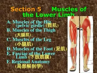
局部解剖学 (二). 股前内侧区 小腿前外侧区. 一、股前内侧区解剖. 浅层结构 深层结构. 皮肤 浅筋膜 浅筋膜内结构 1. great saphenous vein 起始、行程、瓣膜、交通关系、属支、流注. (一)浅层结构. 大隐静脉上段属支的类型. 2. 浅 动 脉. 旋髂浅动脉 superficial iliac circumflex artery 腹壁浅动脉 superficial epigastric artery 阴部外动脉 external pudendal artery.


局部解剖学 (二)
E N D
Presentation Transcript
局部解剖学(二) • 股前内侧区 • 小腿前外侧区
一、股前内侧区解剖 • 浅层结构 • 深层结构
皮肤 • 浅筋膜 • 浅筋膜内结构 1. great saphenous vein 起始、行程、瓣膜、交通关系、属支、流注 (一)浅层结构
2. 浅 动 脉 • 旋髂浅动脉superficial iliac circumflex artery • 腹壁浅动脉superficial epigastric artery • 阴部外动脉external pudendal artery
3. 腹股沟浅淋巴结superficial inguinal nodes 位置、分群、收集区、流注
4. 皮神经cutaneous nerve 1)股外侧皮神经 lateral femoral cutaneous nerve 2)股神经前皮支 anterior cutaneous branches of femoral nerve 3)股神经内侧皮支 medial cutaneous branches of femoral nerve 4)闭孔神经皮支 cutaneous branches of obturator nerve 5)髂腹股沟神经 ilioinguinal nerve 6)生殖股神经股支 femoral branch of genitofemoral nerve
皮神经分布模式图 缝匠肌 • L.C. 股外侧皮神经 • I.C. 股中间皮神经 • M.C. 股内侧皮神经 • OBT. 闭孔神经 • I ING 髂腹股沟神经 • GEN.FEM 生殖股神经的股支
(二)深层结构 Saphenous Hiatus 隐静脉裂孔 1. 股前区深层结构 • 大腿深筋膜deep fascia of thigh (阔筋膜 fascia lata) • 厚而致密 • 髂胫束 iliotibial tract • 隐静脉裂孔saphenous hiatus • 股内侧、外侧、后肌间隔 隐股点
Intermuscular Septum and Osseofascial Sheath of Thigh Anterior osseofascial sheath Medial intermuscular septum Lateral intermuscular septum Medial osseofascial sheath posterior intermuscular septum Posterior osseofascial sheath • 骨筋膜鞘的构成及其内容
肌 腔 隙 lacuna musculorum血管腔隙 lacuna vasorum • 构成与内容 腔隙韧带 髂耻韧带 腹股沟韧带
股 鞘femoral sheath股 管femoral canal • 定义 • 形态 • 内容
股三角femoral triangle • 位置 • 境界 • 内容物及其排列
收肌管adductor canal • 位置 • 境界 • 内容物及其排列
股动脉 femoral artery • 股深动脉 deep femoral artery • 旋股内侧动脉 medial circumflex femoral artery • 旋股外侧动脉 lateral circumflex femoral artery • 穿动脉 perforating arteries
股神经femoral nerve • 起源 • 位置 • 分支 • 分布
2. 股内侧区 • 股内收肌群 耻骨肌 长收肌 大收肌 短收肌 股薄肌 • 闭孔血管神经束 闭孔动脉obturator a. 闭孔静脉obturator v. 闭孔神经obturator n.
二、小腿前外侧区 • 浅层结构 • 深层结构
(一)浅层结构 1. 皮肤 2. 浅筋膜 3. 浅筋膜内结构 • 大隐静脉 Great saphenous vein • 隐神经 Saphenous nerve • 腓浅神经 Superficial peroneal n. • 小隐静脉 Small saphenous vein • 足背静脉弓 Dorsal venous arch • 腓肠神经 Sural nerve
(二)深层结构 • 深筋膜
小腿前、后肌间隔 • 小腿前、外侧、后骨筋膜鞘(构成与内容)
小腿前骨筋膜鞘anterior osseofascial sheath of leg • 小腿前群肌 • 胫骨前肌 Tibialis anterior • 拇长伸肌 Extensor hallucis longus • 趾长伸肌 Extensor digitorum longus • 血管神经束 • 胫前动脉 Anterior tibial artery • 胫前静脉 Anterior tibial vein • 腓深神经 Deep peroneal nerve
小腿外侧骨筋膜鞘lateral osseofascial sheath of leg • 小腿外侧群肌 • 腓骨长肌 Peroneus longus • 腓骨短肌Peroneus brevis • 血管神经束 • 腓浅神经Superficial peroneal nerve
三、足 背 • 浅层结构 1. 皮肤 2. 浅筋膜 3. 浅筋膜内结构 • 大隐静脉 Great saphenous vein • 隐神经 Saphenous nerve • 腓浅神经 Superficial peroneal n. • 小隐静脉 Small saphenous vein • 足背静脉弓 Dorsal venous arch • 腓肠神经 Sural nerve
深层结构 伸肌上支持带 1. 深筋膜 伸肌上支持带 superior extensor retinaculum 伸肌下支持带 inferior extensor retinaculum 骨纤维管 osseofibrous tunnel 2. 足背外来肌: 胫骨前肌腱、拇长伸肌腱、 趾长伸肌腱、第三腓骨肌 3. 足背肌: 拇短伸肌、趾短伸肌 伸肌下支持带
腓骨上支持带 superior peroneal retinaculum 腓骨下支持带 inferior peroneal retinaculum
足背的血管神经 腓深神经 deep peroneal nerve 足背动脉 dorsal artery of foot
操 作 步 骤 (一)股前内侧区解剖 1.去除浅筋膜,保留浅血管、皮神经 2.去除阔筋膜,保留髂胫束。 3.修洁股前群肌:股四头肌、缝匠肌 4.解剖股三角及其内容 (1)观察股三角的边界 (2)在股鞘前壁作三个纵行切口,显露其腔内的股动脉、股静脉和股管。探察股管内容物和股环的边界。
5.解剖收肌管及其内容 在缝匠肌的肌支下方横断此肌,纵行剪开收肌腱板,修洁收肌管内的隐神经、股动脉、股静脉等。 6.解剖股动脉及其分支(股深动脉、旋股内侧动脉和旋股外侧动脉、穿动脉) 7.解剖股静脉及其属支 8.解剖股神经及其分支 9.解剖股内侧群肌:修洁耻骨肌、长收肌、大收肌、股薄肌;横断长收肌,暴露其深面的短收肌;修洁短收肌浅面的闭孔血管、神经前支;横断短收肌,找到其深面的闭孔血管、神经后支
(二)小腿前区 1.去除浅筋膜,保留浅血管、皮神经。 2.去除小腿深筋膜,保留上、下伸肌支持带。 3.解剖小腿前群肌:修洁胫骨前肌、拇长伸肌、趾长伸肌和第三腓骨肌。 4.解剖胫前血管和腓深神经:在小腿上部,钝性分离胫骨前肌与趾长伸肌,在两肌之间、小腿骨间膜前方寻找并清理胫前血管和腓深神经;在小腿下部,钝性分离胫骨前肌与拇长伸肌,在两肌之间、小腿骨间膜前方寻找并清理胫前血管和腓深神经。
(三)小腿外侧区 1.去除浅筋膜,保留浅血管、皮神经。 2.去除小腿深筋膜,保留腓骨上、下支持带。 3.解剖小腿外侧群肌:修洁腓骨长、短肌。 4.解剖腓总神经及其分支:在腓骨颈外侧找出腓总神经;用止血钳沿腓总神经方向向前插入腓骨长肌内,沿腓总神经的走向,切断该肌,显露腓总神经及其分支;追踪腓浅神经和腓深神经达足背。
(四)足 背 1.去除浅筋膜,保留浅血管、皮神经。 2.去除足背深筋膜,保留上、下伸肌支持带。 3.显示骨纤维管:正对胫骨前肌腱、母长伸肌腱和趾长伸肌腱,分别纵行切开伸肌上、下支持带,检查其骨纤维管内肌腱及其腱鞘和血管神经束。 4.解剖足背的肌肉:清理母长伸肌腱、趾长伸肌腱、母短伸肌和趾短伸肌。 5.解剖足背动脉、足背静脉和腓深神经:在母长伸肌腱与趾长伸肌腱之间找到血管神经,并追踪到第2跖间隙近侧端,寻找足背动脉的分支——第1趾背动脉和足底深支。