Essential Guide to Enamel Structure and Composition in Oral Pathology
780 likes | 1.11k Vues
Explore the physical and chemical properties, structure, and submicroscopic features of enamel in dental pathology. Learn about enamel rods, submicroscopic structure, optical phenomena, and striae of Retzius.

Essential Guide to Enamel Structure and Composition in Oral Pathology
E N D
Presentation Transcript
ENAMEL Dr Riaz Abdulla Professor Dept. of Oral Pathology
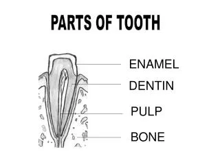
ENAMEL-Physical properties • Hardest tissue in the human body • Extremely hard ( 343 KHN) and brittle • Colour ranges from yellowish white to greyish white • Translucency related to degree of calcification • Has permeability
Does not repair or regenerate • Ameloblasts ( cells of Enamel) are Ectodermal in origin • Thickness • Refractive index- 1.62 • Specific gravity 2.8 • Soluble in acids
ENAMEL- Chemical composition • Inorganic– 96% carbanoapatite crystals (hydroxyapatite substituted with carbonate ions • Organic-4%-Non collagenousprotiens Enamel proteins Low mol wt amelogenins (90%)- (TRAP) Non amelogenins(10%)- enamelin,ameloblastin,tuftelin
ENAMEL ROD • Fundamental organizational unit • Roughly cylindrical in shape with a body(head) and a tail • Average diameter is 4 micron
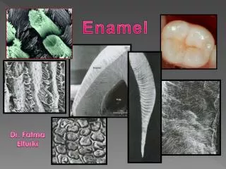
Under the light microscope enamel is composed of roughly cylindrical enamel rods embedded in interrod enamel
Light microscopy • Enamel rods are hexagonal/round/oval in cross section • Appearance of enamel rods surrounded by rod sheath and separated by inter rod substance is the common pattern often referred to as keyhole shaped enamel prisms
Under the light microscope enamel is composed of roughly cylindrical rods embedded in interrod enamel
Enamel-Submicroscopic structure • Structure of enamel rod is beyond the resolution limit of the light microscope • Enamel rods are composed of closely packed and long ribbon like hydroxyapatite crystals 600-1000 A long and 250 A thick. • The roughly cylindrical shaped enamel rods are made up of crystals that run parallel to the long axis in the central part. • Crystals away from the central axis of the rod flare laterally
Inter rod region • Surrounds each rod • Crystals oriented in a different direction except in the interrod region located directly cervical to a particular rod
Rod sheath • Narrow space /boundary between rod and interrod enamel and containing organic material • Rod sheath is the boundary where crystals of rod meet those of interrod region at sharp angles
Direction of enamel rods • At right angles to dentin surface/DEJ • Horizontal in cervical and central part of deciduous teeth • Apically directed in cervical portion of permanent teeth • Enamel rods at pit and fissure region of occlusal surface of premolars and molars converge in their outer course (lateral flaring towards DEJ)
Enamel stuctures due to optical phenomenon • GNARLED ENAMEL • HUNTER SCHREGER BANDS
Gnarled Enamel • Enamel rods at the region of cusps/incisal edges seem to intertwine irregularly • The interwining and twisting of enamel rods gives rise to a special optical appearance called gnarled enamel
GNARLED ENAMEL • Twisting / Undulation / Overlapping of enamel rods. • Cuspal & Incisal • Optical
ImportanceTwisting of rods Increases strength Resist the load •Reason:- Ameloblast retreat in a very irregular course initially
Hunter Schreger bands • Alternating light and dark bands seen in longitudinal sections under oblique reflected light • Optical phenomenon caused by total internal reflection • Variations in calcification,organic content permeability etc accounts for it
Dark zones - Diazone Light zones Parazone
Hypocalcified structures •Incremental lines (Striae) of Retzius •Enamel spindle •Enamel tufts •Enamel lamellae
Striae of Retzius • Brownish bands in ground sections of enamel • Incremental pattern of formation • Seen at right angles to enamel rods
•Appositional process •Alternating periods of growth & rests •2 Types 1.Major- Incremental lines of Retzius 2.Minor - Cross striations
Cross striations •Irregular lines crossing at regular intervals over enamel rods •Perpendicular to long axis of the rod •Daily increments •4 μ apart (secretion of 1 day)
Striae of Retzius •Major •Striae – “ THICK” •Irregularly spaced •Dark brown •Oblique •From DEJ to outer surface
20-80 μ m apart • Width – 4-15 μ • Deposition 7-14 days • Distance b/w lines • More – Occlusal / Incisal • Less - Cervical • Hypo calcified areas • Prominent – Permanent teeth
Reasons ???? • Difference in Mineralization • Change in rod direction • Change in crystal orientation • Defective enamel matrix production
Types of striae 1.Complete 2.Incomplete Complete striae- • Does not reach the surface • Encircle the dentin tip • Cuspal & Incisal • Cross section- Concentric circles
Incomplete striae- • Reach the surface • Results in Perikymata
Cross striations Daily variation (Circadian rythm) • •Striae of retzius Weekly rythm
Perikymata • Transverse Wave like grooves • External manifestation of Striae of retzius • Newly erupted teeth • Disappears - AGE