Download
1 / 39
410 likes | 686 Vues
p 53 Missense Mutation C ancer. Outline. Disease related to p53 Role and regulation pathway Structure of p53 Missense mutation and consequences E xperiment’s proposal of p53 missense mutant cancer. I. Diseases cause by mutated p53. Aneuploidy phenotype Li- Fraumeni syndrome (rare)

E N D
Outline • Disease related to p53 • Roleand regulation pathway • Structure of p53 • Missense mutation and consequences • Experiment’s proposal of p53 missense mutant cancer
I. Diseases cause by mutated p53 • Aneuploidy phenotype • Li-Fraumenisyndrome (rare) • 50% of human cancers • Breast and colonrectum cancers • Bladder cancers • Lung cancer • Hypoxia • Glioblastomamultiforme (GBM)
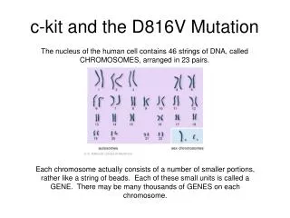
TP53 Gene • TP53 encodes p53 protein, located on short arm of chromosome 17 at position 13.1
Roles of p53 • p53 before was oncogene • “Guardian of the genome” • p53 gene today—tumor suppressor or anticancer • P53 functions at all checkpoints during the cell cycle
Roles of p53 • Functions as transcription factor
Regulations of p53 • P53 has very complicated pathway and network as the tumor suppressor gene • Activation state: 1. upstream pathways 2. downstream pathways • Inactivation state
Active state • p53 is always in an off stage • Only activate when cells are stressed or damaged
1. Upstream Regulation of p53 In stressed conditions, p53 is activated by:
2. Downstream Activation of p53 The turn on of p53 lead to: • cell-cycle regulation, • initiate growth arrest • induction of apoptosis • control development and differentiation
Inactivation state • In unstressed condition, p53 is inactivate by Mdm2-binding • Mdm2 is an ubiquitin ligase
Concentration Level of p53 • P53 is always in an off stage • Activation of p53 always results in apoptotic and cell cycle arrest • Apoptosis induced and leads to increase level of p53 • MDM2 control the level of p53 creating negative feedback loop • MDM2 is an E3 ligase that targets p53 to ubiquitination
Structures of p53 • Same protein family of p63 and p73 • P53,p63, p73 family of transcription factor • A nuclear phosphoprotein of MW 53kDa • Open reading frame consists of 393 amino acids • Has 7 domains • The central part is where DNA contacts with the protein
N-teminus(TAD) • 1. Transactivation • 2. Activation domain 2 (AD2) • 3.Proline rich region • Central core • 4. DNA-binding core domain (DBD)– has Zn and several Arginine amino acids • C-terminus • A. Tetramerization: • 5. Nuclear localization signaling domain (NLS) • 6. Homo-oligmerisation domain (OD) • B. Down regulation of DNA binding: • 7. regulatory domain
Crystal structure of p53 at Core Domain The DNA binding domain is folding as beta barrel or beta-sandwich
Structure of p53 • p53 forms a tetramer : • _ 4 helical bundle fold • _essentials for DNA binding, protein-protein interactions, post translational modifications, and p53 degradation • _oligomerisationdomain consists of a dimer of dimers
p53 Missense Mutation • Majority of p53 mutation occurs at DNA binding core • >50% occurs at single missense mutation • Hydrophobic and unstable • Loss of stability and loss of molecular contact inactivate p53 • Destabilize and deformed p53
Residues in Unstable MutationTable 2. Fractions of unstable mutants (>3kT) from codons 96-289 before and after substitution
Residues in Unstable Missense Mutation • Common mutated residues are R248 and R273. These two residue directly binding to DNA ligand. • R175, R248, and R282 are help stabilizing the structure of DNA binding interface.
Significant of p53 to Cancer Treatment • Target the protein and the pathways of the protein might rescue p53 and restore its functions.
PURPOSE OF THE EXPERIMENT 1st Goal: Design the input files and the starting structures for p53 cancer mutants. 2nd Goal: Using the AMBER software to run the stimulation and to compare the binding affinity between the mutations and the native states of p53, either with or without DNA ligand.
Hypothesis • Missense mutation due to the destabilize and deformation in the DNA binding domain (DBD) deactivate p53, affecting protein-ligand interaction. • Missense mutated at R248, R249 and R282 position will prevent the transcription activation and destroy the ability of p53 to bind to its target DNA sequence.
Methods • Downloading the crystal structure of 1TRS as native state of p53 protein • Creating and designing a series of input files (prmtop and inpcrd) to run the stimulations for the both the native state and the destabilized cancer mutants at the sites of R248, R249 and R282 • Compare the stimulated stabilities between these two mutants with and without DNA binding by using MM-PBSA in the Amber software CHAIN B
Function of p53 • P53 has many anticancer mechanisms, and plays a role in apoptosis, genetic stability, and inhibition of angiogenesis. In its anti-cancer role, p53 works through several mechanisms: • It can activate DNA repair proteins when DNA has sustained damage. • It can induce growth arrest by holding the cell cycle at the G1/S regulation point on DNA damage recognition (if it holds the cell here for long enough, the DNA repair proteins will have time to fix the damage and the cell will be allowed to continue the cell cycle). • It can initiate apoptosis, the programmed cell death, if DNA damage proves to be irreparable. • Activated p53 binds DNA and activates expression of several genes including WAF1/CIP1 encoding for p21. p21 (WAF1) binds to the G1-S/CDK (CDK2) and S/CDK complexes (molecules important for the G1/s transition in the cell cycle) inhibiting their activity.
Diseases Loss of p53 creates genomic instability that most often results in the aneuploidy phenotype.[28] Li-Fraumeni syndrome (rare) the mutation or loss of the p53 gene can be identified in more than 50% of all human cancer cases worldwide.
Structure of p53 • Encode by TP53 gene, a 20 Kb gene that contains 11 exons and 10 introns • Located on the small arm of chromosome 17 • Highly conserved gene