Download
1 / 37
370 likes | 1.76k Vues
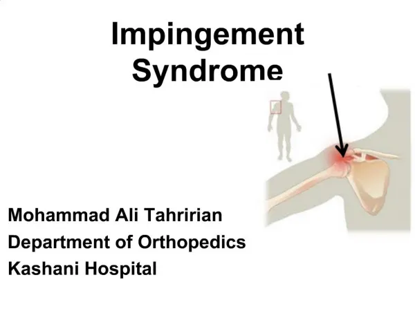
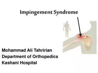
SHOULDER IMPINGEMENT SYNDROME. Definition: Shoulder impingement has been defined as compression and mechanical abrasion of the supraspinatus as they pass beneath the coracoacromial arch during elevation of the arm. Related terms:

E N D
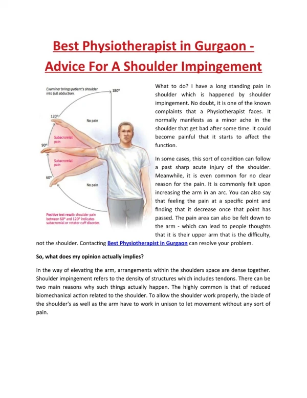
Definition: Shoulder impingement has been defined as compression and mechanical abrasion of the supraspinatus as they pass beneath the coracoacromial arch during elevation of the arm. Related terms: Rotator cuff tendinitis: It encompasses impingement, and result from acute rotator cuff overload, intrinsic rotator cuff degeneration, or chronic overuse. Rotator cuff syndrome: It is a term used to describe the process whereby tendinitis and impingement are ongoing simultaneously. Painful arc syndrome: Pain in the shoulder and upper arm during the midrange of glenohumeral abduction, with freedom from pain at extremes of the range due to supraspinatus damage . The term shoulder impingement syndrome has largely replaced what used to be called painful arc syndrome.
Functional anatomy: The rotator cuff (Figure 21) comprises four muscles The subscapularis, the supraspinatus, the infraspinatus and the teres minor and their musculotendinous attachments. The subscapularis muscle is innervated by the subscapular nerve and originates on the scapula. It inserts on the lesser tuberosity of the humerus. The supraspinatus and infraspinatus are both innervated by the suprascapular nerve, originate in the scapula and insert on the greater tuberosity.
The teres minor is innervated by the axillary nerve, originates on the scapula and inserts on the greater tuberosity. A bursa in the subacromial space provides lubrication for the rotator cuff.
The rotator cuff is the dynamic stabilizer of the glenohumeral joint. The static stabilizers are the capsule and the labrum complex, including the glenohumeral ligaments. Although the rotator cuff muscles generate torque, they also depress the humeral head. The deltoid abducts the shoulder. Without an intact rotator cuff, particularly during the first 60 degrees of humeral elevation, the unopposed deltoid would cause cephalic migration of the humeral head, with resulting subacromial impingement of the rotator cuff. In patients with large rotator cuff tears, the humeral head is poorly depressed and can migrate cephalad during active elevation of the arm.
Etiology: 1. Extrinsic causes: A- Bony factors: • The type I acromion, which is flat, is the "normal" acromion. • The type II acromion is more curved and downward dipping, • The type III acromion is hooked and downward dipping, obstructing the outlet for the supraspinatus tendon and therefore may impinge on the rotator cuff on elevation of the arm. • Osteophytesunder the acromioclavicular joint reduces the subacromial space and can also lead to cuff impingement and therefore failure" '
Type I Type II Type IIIFigure 22 : Types of anatomical acromion variation: Flat acromion, curved and hoocked
B- Soft tissue factors Examples include • • Subacromial bursitis • • Thickened coracoacromial ligament. 2. Intrinsic causes a. Degenerative cuff failure : This constitutes the commonest cause of cuff failure and usually occurs in the older individual. Degeneration of the cuff may later result in partial tears which may progress to complete tears. The precise cause of degenerative cuff tear is unknown. One possible theory relates to the 'critical vascular zone' of the cuff tendon where the blood supply is precarious, and relative ischemia leads to degenerative changes. b. Traumatic cuff failure: This may occur when the upper limb is subject to a violent force and the rotator cuff sustains a traumatic tear. In the younger individual where the tendinous part of the cuff-bone complex is stronger than the bony part, the tendons may avulse with a piece of bone.
c. Reactive cuff failure: Calcific rotator cuff tendinitis is an example of reactive cuff failure. The calcifying mass inside the tendon may give rise to a swelling which leads to impingement under the subacromial arch, hence resulting in cuff failure.
Classification of the Impingement Syndrome Neer divided impingement syndrome into three stages: 1. Stage Iinvolves edema and/or hemorrhage. This stage generally occurs in patients less than 25 years of age and is frequently associated with an overuse injury. Generally, at this stage the syndrome is reversible. 2. Stage II is more advanced and tends to occur in patients 25 to 40 years of age. The pathologic changes that are now evident show fibrosis as well as irreversible tendon changes. 3. Stage III generally occurs in patients over 50 years of age and frequently involves a tendon rupture or tear.
History : 1- Pain:It is exacerbated by overhead or above the shoulder activities. A frequent complaint is night pain, often disturbing sleep, particularly when the patient lies on the affected shoulder. The onset of symptoms may be acute, following an injury, or insidious, particularly in older patients, where no specific injury occurs. In the acute stage I, there is a painful arc of abduction between 60 and 120 degrees increased with resistance at 90 degrees. 2- Loss of motion : Prolonged shoulder pain causes the patient to restrict instinctively the range of use and often results in an initial adhesive capsulitis. 3- weakness and inability to raise the arm may indicate that the rotator cuff tendons are actually torn.
Physical examination: 1. Manual motor testing for the rotator cuff muscles: Geber's lift-off test for subscapularis External rotation with adducted and elbow flexed 90 degrees for test of the infraspinatus and teres minor. Arm abduction 90 degrees in the scapular plane (30 degrees anterior to the coronal plane of the body and internal rotation for test of the supraspinatus.
Figure 23: Lift off test for subscapularis, external rotation for teres minor and infraspinatus and abduction with internal rotation for supraspinatus test
2. The key feature of the physical examination is an assessment for signs of impingement: a-Neer impingement sign: With the patient seated or standing place one hand on the posterior aspect of the scapula to stabilize the shoulder girdle, and, with the other hand, take the patient's internally rotated arm by the wrist, and place it in full forward flexion. If there is impingement, the patient will report pain in the range of 70 degrees to 120 degrees of forward flexion as the rotator cuff comes into contact with the rigid coracoacromial arch.
b-Hawkins impingement sign: With the patient sitting or standing, the examiner places the patient's arm in 90 degrees of forward flexion and forcefully internally rotates the arm, bringing the greater tuberosity in contact with the lateral acromion. A positive result is indicated if pain is reproduced during the forced internal rotation at the supraspinatus site. C-AROM of shoulder : Forward flexion, abduction, external rotation and internal rotation.
Figure 26: AROM of shoulder flexion, abduction, ext. rotation with 90 abduction and neutral the last is Apleys scratch test for internal rotation.
Management: There are three ways of approaching impingement syndrome: І-Physical therapy rehabilitation, ІІ-subacromial injections of cortisone, and ІІІ-surgical intervention. І -Physical therapy rehabilitation in : 1- Pain control and inflammation reduction by: • Relative rest: A sling may be used but it is crucial that the sling be removed several times daily to perform exercises. Acute phase
Icing (20 min, 3-4 times per day): It decreases the size of blood vessels in the sore area. • Have the patient sleep with a pillow between the trunk and arm to decrease tension on the; upraspinatus tendon (that is the arm is littleabduction, flexion and internal rotation) and prevent blood flow comprise in its watershed region. • Patients are instructed to continue the pain control techniquesat home, work, or vacation as part of their exercise program. The home exercise program builds on itself through each phase of the rehabilitation process, and carry-over should be monitored
Recovery Phase The recovery phase from a rotator cuff injury must include several components to be successful. These include the following: • Restoration of shoulder ROM, • Normalization of strength and dynamic muscle control, and • Proprioception and dynamic joint stabilization.
1-Restoration of shoulder range of motion After the pain has been managed, restoration of motion can be initiated: • Codman pendulum exercises. • Wall walking • Stick or towel exercises • Address any posterior capsular tightness because this can lead to anterior and superior humeral head migration, resulting in impingement: Stretching of the posterior capsule. The focus of treatment in this early stage should be on improving range, flexibility of the posterior capsular postural biomechanics, and restoring normal scapular motion. Each stretch should be held for a minimum of 30 seconds, although stretching for 1 minute is encouraged.
2-Normalization of strength and dynamic muscle control a.Perform strengthening in a pain-free range only. Begin with the Scapulothroracic stabilizers to help return smooth motion allowing normal rhythm between scapula and GH joint. The scapular stabilizers include the rhomboids, levator scapulae, trapezius, and serratus anterior.: • Shoulder shrugs. • push-ups. b.Then, turn attention toward strengthening the rotator cuff muscles. Position the arm at 45° and 90° of abduction for exercises to prevent the wringing out phenomenon, in which hyperadductioncan be caused, stressing the tenuous blood supply to the tendon of the exercising muscle. Avoid the thumbs down position with the arm in greater than 90° of abduction and internal rotation to minimize subacromial impingement.
Many ways to strengthen muscles are available. The rehabilitation program usually starts with isometricprogresses to concentric contractions, and finally incorporates eccentric contractions as part of the preparation for return to sports. Additional strengthening techniques that can be used are progressive resistive exercises (PREs), Thera-Band, and plyometrics.Use of isokinetic exercises has been debated because they are not performed in a functional manner. Probably the best use for isokinetic exercise machines is for objective side-to-side comparison of strength and progress made in strength rehabilitation. Incorporate endurance training into the program as it advances.
Stick exerciseFigure 27: Shoulder stretching exercises include gentle pendulum exercises, stick exercises, the use of overhead pulley.
Figure 28:The shoulder strengthening program is designed to improve strength in the remaining rotator cuff and improved strength of the deltoid. The five theraband exercises provide resistance against internal rotation and external rotation , abduction, adduction, extension and forward flexion to strengthen the rotator cuff muscles and the three distinct portions of the deltoid muscle.
Shoulder press up upsFigure 29: Scapular stabilizer are strengthened by shoulder shrug, push-up and shoulder press
3-Proprioception Proprioceptive training is important to retrain neurologic control of the strengthened muscles, providing improved dynamic interaction and coupled execution of tasks for harmonious movement of the shoulder and arm. Begin tasks with closed kinetic chain exercises to provide joint stabilizing forces. Then as the muscles become reeducated, one can progress to open chain activities, In addition, proprioceptive neuromuscular facilitation (PNF) is designed to stimulate muscle/tendon stretch receptors for reeducation.
Maintenance Phase Return to task-specific or sport-specific activities is the last phase of rehabilitation. This phase is an advanced form of proprioceptive training for the muscles to relearn prior activities. It is an important phase of rehabilitation and should be supervised properly to minimize the possibility of re injury. At the conclusion of formal therapy sessions, patients should be independent in a ROM and strengthening program and should continue these exercises. Athletes are often tempted to return to their overhead throwing sport too soon after recovery of the acute phase.
ІІ-Subacromial injections of cortisone: Although these injection do not cure the underlying pathology, they decrease swelling of the inflamed bursa and rotator cuff tissue and allow for more room in the sudacromial space for the rotator cuff to move. Corticosteroidsdelivered directly to the subacromial space via injection can be considered.
ІІІ -Surgical Intervention Indications for operative treatment of rotator cuff disease include partial-thickness or full-thickness tears in an active individual who does not improve pain and/or function within 3-6 months with a supervised rehabilitation program. An acromioplasty is usually performed in the presence of a type II (curved) or type III (hooked) acromion with an associated rotator cuff tear. In surgical candidates, early repair is useful to avoid fatty degeneration and retraction of the remnant rotator cuff musculature