Download
1 / 43
440 likes | 772 Vues
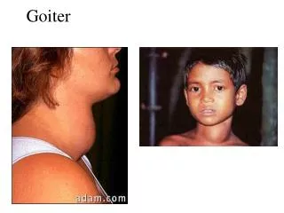
Goiter. * Definition: Non-inflammatory, non-neoplastic enlargement of the thyroid gland. * Classification: Simple (non-toxic) goiter. Toxic goiter. SIMPLE (NON-TOXIC) GOITER. Enlargement of the thyroid without toxic manifestations. * Causes: 1. Iodine deficiency.

E N D
* Definition:Non-inflammatory, non-neoplastic enlargement of the thyroid gland. * Classification: • Simple (non-toxic) goiter. • Toxic goiter.
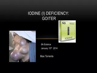
SIMPLE (NON-TOXIC) GOITER • Enlargement of the thyroid without toxic manifestations. * Causes: 1. Iodine deficiency. a. Absolute deficiency: in areas far from the sea. b. Relative deficiency: due to increased demand for iodine at pregnancy, puberty and lactation. 2. Dyshormonogenesis: hereditary deficiency of enzymes necessary for thyroxine formation. 3. Goitrogens: Well-known goitrogens as cabbage, cauliflower which contain thiocyanate which inhibits iodide transport within the thyroid.
* Pathogenesis: a. Parenchymatous goiter: • Iodine deficiency → decreased thyroid hormone synthesis → increases TSH secretion → thyroid glands hyperplasia. • The acini are increased in number and lined by tall columnar cells and contain little colloid. • If iodine deficiency is corrected after a short time, the acini return to the normal state.
b. Colloid goiter: • When iodine deficiency is corrected after a longer time → the acini are distended with colloid and lined by flat cells. c. Nodular goiter: • Repeated cycles of iodine deficiency & correction →nodular goiter in which the gland shows multiple nodules of parenchymatous goiter, colloid goiter and areas of fibrosis.
* Morphological features: a. Parenchymatous goiter: * Gross picture: • Symmetrical enlargement. • Firm in consistency. • Cut surface is grayish pink.
* Microscopic picture: • Hyperplastic acini lined by tall columnar cells and filled with scanty colloid.
b. Colloid goiter: * Gross features: • Symmetrical enlargement. • Soft in consistency. • Cut surface is grayish brown in color and may shows cystic spaces filled with glistening colloid “honey comb appearance”.
* Microscopic picture: • The acini are distended with colloid and lined by flat cells. • The stroma is scanty.
c. Nodular goiter: * Gross picture: • Asymmetrical enlargement. • Variable: firm areas and soft cystic areas. • Cut surface is nodular.
* Microscopic picture: • Multiple nodules, some formed of hyperplastic acini and others show acini filled with colloid. • The nodules are surrounded by fibrous tissue.
* Complications of simple goiter: 1. Pressure effects: on esophagus, trachea, and recurrent laryngeal nerve. 2. Secondary hyperthyroidism due to Hyperfunctioning nodules (toxic nodular goiter). No exophthalmos. 3. Malignancy in 2% of cases: follicular carcinoma.
Toxic goiter • Two types; 1. Primary toxic goiter (exophthalmoic goiter or grave’s disease). 2. Secondary toxic goiter: toxic nodular goiter or toxic adenoma.
Organ specific autoimmune disease due to auto-antibodies (LATS; long acting thyroid stimulating) stimulating TSH receptors leads to diffuse hyperplasia and hyperfunctioning acini with excess thyroid hormone secretion
* Pathological features: 1. Thyroid: • N/E: symmetrically enlarged, firm, with dark red “vascular” cut surface. • M/P: hyperplastic acini lined by columnar cells and filled with faintly stained colloid with peripheral scalloping. The stroma is highly vascular and shows lymphocytic infiltration.
Graves’ disease :Diffusely enlarged gland , Can weigh up to 200 g ,Richly vascular
Toxic goiter:scalloping of colloid inside thyroid folliclessmall sized follicles , lymphocytic infiltration,hypervascularity
2. Exophthalmos:forward protrusion of the eye globe due to edema and degeneration of the retro-orbital muscles “special auto-antibodies react with them”. 3. Diffuse lymphoid hyperplasia: in thymus, tonsil, spleen, guts. 4. Left ventricular hypertrophy “thyrotoxic cardiomyopathy”. 5. Pre-tibial myxedema. 6. Increased basal metabolic rate
* Causes: A. Toxic nodular goiter: • Complicating simple nodular goiter. • Diffuse, nodular enlargement of the thyroid. Some nodules show hyperfunctioning acini. Other acini are inactive. B. Toxic adenoma: • Complicating thyroid adenoma. • The Hyperfunctioning neoplastic acini are like those of grave’s disease. The remaining thyroid tissue is inactive. Thyroid hormone secretion is autonomous.
Inflammation of the thyroid. * Types: 1. Hashimoto’s thyroiditis. 2. Subacute granulomatous thyroiditis (DeQuervain thyroiditis). 3. Reidel’s (fibrous) thyroiditis.
hashimoto thyroiditis • Occurs in middle & old age. • Common in females more than males (20:1). • Cause painless thyroid enlargement. • Associated with hypothyroidism. * Pathogenesis: • Autoimmune disease in which the immune system reacts against a variety of thyroid antigens.
* Gross picture: • Symmetrically enlarged thyroid gland. • Firm inconsistency. • Intact, non-adherent capsule. • Cut surface is pale, homogenous and sometimes nodular.
* Microscopic picture: • Dense inflammatory infiltrate formed of lymphocytes, plasma cells and macrophages, with sometimes lymphoid follicle formation. • Some acini are atrophied and others show regenerative changes (lined by large cubical cells with deeply esinophilic granular cytoplasm termed (Hurthle cells). This is termed Hurthlecell metaplasia. • Finally, fibrosis.
* Complications: • Hypothyroidism. • Development of other autoimmune diseases. • Malignant transformation (lymphoma)
Subacute granulomatous thyroiditis • Occurs between 30-50 years. • More common in females than males (5:1). • Cause painful thyroid enlargement. • Associated with transient hyperthyroidism. * Pathogenesis: • Associated with viral infection.
* Gross picture: • Unilateral or bilateral enlargement. • Intact capsule. • Slightly adherent. • Cut surface shows scattered firm yellowish white areas.
* Microscopic picture: • Neutrophilic infiltration with variable destruction of the thyroid follicles. • Pools of colloid surrounded by multinucleate giant cells, aggregations of lymphocytes, histiocytes and plasma cells. • Finally, fibrosis, chronic inflammatory cells replace the damaged foci.
Reidel’s thyroiditis • Rare, of unknown cause. Affect both sexes equally. * Gross picture: • The gland is hard in consistency and adherent to the surrounding structures (simulating malignancy). * Microscopic picture: • Dense fibrous tissue replacing the thyroid tissue and penetrating the capsule to the surrounding neck structures.