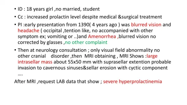
Understanding the Lymphatic System: Structure, Functions, and Components
540 likes | 664 Vues
The lymphatic system is a vital vascular network responsible for returning excess tissue fluid to the bloodstream. Composed of lymphatic vessels, tissues, and organs, it plays key roles in immune responses, production of lymph cells, destruction of pathogens, and the clearance of foreign particles. Lymphatic vessels, classified into capillaries, trunks, and ducts, are essential for fluid circulation. This system, distinct from the cardiovascular system, ultimately connects to the venous system, helping maintain fluid balance and support immunity across the body.

Understanding the Lymphatic System: Structure, Functions, and Components
E N D
Presentation Transcript
No. 18 • The Lymphatic System
Section 1 Introduction • The lymphatic system is a separate vascular system serving as accessory system for the return of fluid from the tissue spaces. • The fluid in the lymphatic vessels is termed lymph. Its composition is similar to that of plasma, except for the lower concentration of proteins. • This system does not open into the heart but into the venous system in the root of the neck.
1. Constitution of the lymphatic system • The lymphatic system consists of the lymphatic vessels, lymphatic tissue, and lymphatic organs.
2. functions of the lymphatic system • The lymphatic system has several important functions. • The functions of the lymphatic system include the producing lymph cells, the destruction of bacteria, the removal of foreign particles from the lymph, specific immune responses, and the return of interstitial fluid to the bloodstream.
Ⅰ The Lymphatic Vessels • The are classified according to size and structure as lymphatic capillaries, lymphatic vessels, lymphatic trunks and lymphatic ducts.
Ⅰ) Lymphatic capillary • The lymphatic capillariesform network in the tissue spaces, the meshes of which are larger than those of the neighbouring blood capillaries. They often commence with a dilated, bulb-like, blind end and their calibre is greater than that of blood capillaries. • The wall is composed of a single layer of overlapping endothelial cells attached by anchoring filaments to the surrounding connective tissue.
Another important feature of the endothelial wall of the lymphatic capillaries is that it is permeable to substances of much greater molecular size than those which can pass through the endothelial wall of blood capillaries.
Lymphatic capillaries are present in many tissue of the body, but are absent from avascular structures (epidermis, hair, nails, cornea, and articular and some other cartilages), the brain and spinal cord, the splenic pulp and bone marrow.
Ⅱ) The lymphatic vessels (lymphatics) • The lymphatic vessels are united with lymphatic capillaries. They have three-layered wall similar to the wall of veins. Their valves, which are more numerous than in veins, permit lymph to flow in only one direction and give lymphatic vessels a characteristic beaded appearance. The lymphatic vessels course in close relationship with the veins of any body part. They exceed the veins in number, but they are smaller in size and thinner walled. At intervals along the course of the lymphatic vessels, lymph nodes are interposed.
Likes veins, the lymphatic vessels are arranged in superficial and deep sets. • The superficial lymphatics lie immediately under the skin and may run independently or may accompany the superficial veins; they drain lymph from the superficial structures. The deep lymphatics always accompany vascular or neurovascular bundles; they drain lymph from muscles and internal organs.
Ⅲ) The lymphatic trunks • Having traversed the corresponding nodes, the lymphatic vessels from the various parts of the body unite one another and finally form nine lymphatic trunks, which are named, • The right and left jugular trunks(2), • the right and left subclavian trunks(2), • the right and left bronchomediastinal trunks(2), • the right and left lumbar trunks(2), • and the intestinal trunk(1).
Ⅳ) The lymphatic ducts • There are two lymphatic ducts in number. • The right lymphatic duct is formed by the union of the right jugular, right subclavian and right bronchomediastinal trunks. It opens into the right venous angle • The thoracic duct is formed by the union of the right lumbar, left lumbar and intestinal trunks, at the root of the neck it receives the left jugular, left subclavian and left bronchomediastinal trunks. It opens into the left venous angle.
Ⅱ. The lymphatic Tissue • The lymphatic tissue is situated in the wall of the alimentary, respiratory, urinary and reproductive canals. There are two kinds of lymphatic tissue, that is lymph nodules and scattered lymphatic tissue.
Ⅲ. The Lymphatic Organs • They are lymph nodes, tonsils, spleen and thymus. • The lymph nodes: • They are small, round or bean-shaped organs that are distributed along the course of many of the lymphatic vessels. There are groups of lymph nodes in the groin, axilla, and neck, as well as in numerous other deeper locations. • They may also be divided into the superficial and deep groups.
Each node consists of lymphatic tissue enclosed in a fibrous connective capsule. • Generally each presents on one side a slight depression, termed the hilum, through which the blood vessels enter and leave the node. The efferent lymph vessels (usually single) also emerges from the node at this hilum, while the afferent vessels enter it at several different parts of the periphery. Lymph passes through several groups of lymph nodes before entering the blood stream.
Local corresponding lymph nodes: • Lymph from an area of the body is drained into the local (regional) corresponding lymph nodes. • The lymph nodes can filter out certain amount of microorgainsms and inflammatory products. This process often produces tenderness and swelling in nodes of an infect ed area. If bacteria in an area drained by a node become too numerous, they may attack the node itself, resulting in an abscess.
Lymph node enlargement may be local or widespread. Cause of lymph node enlargement includes infection, allergy, primary disease of the node (such as Hodgkin’s disease, a cancer of the lymph node) and leukemia.
One feature of the lymphatic system is its significance in the spread of tumors. Cancer usually produces a secondary growth in regional lymph nodes. Many of these secondary growths (metastases) result from tumor emboli detaching from the point of origin and lodging in the regional lymph nodes. Operations for the removal of cancer are therefore planned to take away the mass of the cancer, the intervening lymphatic vessels and the lymph nodes.
Section 2 The Lymphatic Ducts • Ⅰ. The Thoracic Duct (left lymphatic duct) • It is the largest lymph vessel of the body and conveys the greater part of the lymph back into the circulating blood. In the adult it varies in length from 30-40 cm.
The duct begins at the upper end of the cisterna chyli near the lower border of the twelfth thoracic vertebra and enters the thorax through the aortic hiatus of the diaphragm. It ascends through the posterior mediastinum with the aorta on its left, the azygos vein on its right side, the vertebral column on its back, and the esophagus on its front. Opposite the fifth thoracic vertebra the thoracic duct passes obliquely behind the esophagus to its left side, and continues superiorly to the thoracic inlet. Then, it arches laterally behind the left carotid sheath at the root of the neck and ends by opening into the left venous angle.
In the neck it is joined usually by the left jugular trunk, the left subclavian trunk and the left bronchomediastinal trunk. • The cisterna chyli is an elongated saccular dilatation in the lymphatic route from the abdomen and lower limbs. It is situated in front of the first and second lumbar vertebral bodies, immediately to the right of the abdominal aorta. It receives the right and left lumbar and intestinal trunks. • The thoracic duct drained the lymph from the lower limbs, pelvis, abdomen, left upper limb, left thorax, and left head and heck, that is the 3/4 parts of the body.
Ⅱ. The right lymphatic duct • It courses along the medial border of scalenus anterior at the root of the neck, about 1 –5 cm long, and ends by opening into the right venous angle. • It is the union of the right jugular trunk, right subclavian trunk, and right bronchomediastinal trunk.
The right lymphatic duct receives the lymph from the right side of the head and neck through the right jugular trunk, from the right upper limb through the right subclavian trunk, from the right side of the thorax, right lung, right side of the heart, and part of the convex surface of the liver through the right bronchomediastinal trunk.
Shortly, it receives the lymph from the right and superior quarter of the body, while the lymph from the rest greater part is collected into the thoracic duct, that is the 1/4 part of the body.
Section 3 The Lymphatic Drainage Ⅰ. The lymphatic vessels and lymph nodes of head and neck Ⅰ) The lymph nodes of head 1. Occipital lymph node 2. retroauricular lymph node 3. parotid lymph node 4. submandibular lymph node 5. submental lymph node
Ⅱ) The lymph nodes of neck 1. Anterior cervical lymph node (1) Superficial anterior cervical lymph node (2) Deep anterior cervical lymph node ① Prelaryngeal lymph node ② Thyroid lymph node ③ Pretracheal lymph node ④ Paratracheal lymph node
2. Lateral cervical lymph node (1) Superficial lateral cervical lymph node (2) Deep lateral cervical lymph node ① Superior deep lateral lymph node ② Inferior deep lateral lymph node • Supraclavicular lymph node • Retropharyngeal lymph node
Ⅱ. The lymphatic vessels and lymph nodes of upper limb Ⅰ) Cubital lymph node Ⅱ) Infraclavicular node Ⅲ) Axillary lymph node 1. Pectoral lymph node 2. Lateral lymph node 3. Subscapular lymph node 4. Central lymph node 5. Apical lymph node
Ⅲ. The lymphatic vessels and lymph nodes of thorax Ⅰ) The lymph nodes thoracic wall 1. Parasternal lymph node 2. Intercostal lymph node 3. Superior phrenic lymph node Ⅱ) The lymph nodes of the thoracic contents 1. Anterior mediastinal lymph node 2. Posterior mediastinal lymph node 3. Tracheobronchial lymph and pulmonary nodes
Ⅳ. The lymphatic vessels and lymph nodes lower limb Ⅰ) Popliteal lymph node Ⅱ) Inguinal lymph node 1. Superficial inguinal lymph node 2. Deep inguinal lymph node
Ⅴ. The lymphatic vessels and lymph nodes of pelvis Ⅰ) Internal iliac lymph node Ⅱ) Sacral lymph node Ⅲ) External iliac lymph node Ⅳ) Common iliac lymph node
Ⅵ. The lymphatic vessels and lymph nodes of abdomen Ⅰ) The lymph nodes of the abdominal wall Lumbar lymph node Ⅱ) The lymph nodes of the abdominal viscera
1. The lymph nodes arranged along the celiac artery and its branches • Celiac lymph nodes ① Right and left gastric lymph nodes ② Right and left gastroepiploic lymph nodes ③ Pyloric lymph nodes ④ Hepatic lymph nodes ⑤ Pancreaticosplenic lymph nodes
2. The lymph nodes arranged along the superior mesenteric artery and its branches • Superior mesenteric lymph nodes ① Mesenteric lymph nodes ② Ileocolic lymph nodes ③ Right colic lymph nodes ④ Middle colic lymph nodes
3. The lymph nodes arranged along the inferior mesenteric artery and its branches • Inferior mesenteric lymph nodes ① Left colic lymph nodes ② Sigmoid lymph nodes ③ Superior rectal lymph nodes
Section 4 The Spleen • In addition to the lymph nodes, several organs are lymphoid in nature. These include the spleen, the thymus gland, and the tonsils. They are named the lymphoid organs. These organs, which have on direct association with the lymphatic system of vessels or with the lymph, are an integral part of the body’s immune system. • The spleenis the largest lymphoid organ.
1. Position • It lies principally in the left hypochondriac region of the abdomen, between the fundus of the stomach and the diaphragm, and its long axis is in line of the tenth rib. • It is not palpable below the left costal arch unless it is enlarged or markedly dislocated. It is soft, of friable consistence, and of dark purplish colour. It is subject to injury from the blunt trauma.
2. Morphology • The spleen has • Two surfaces (diaphragmatic and visceral), • Two borders (superior and inferior), • Two extremities (anterior and posterior). • The diaphragmatic surface is convex, smooth and faces upwards and backwards. It is in relation with the abdominal surface of the diaphragm.