Cell Membranes
Cell Membranes. What ’ s wrong with this picture?. http://www.studyblue.com/notes/note/n/osmolality-and-osmo-gap/deck/1598495. The Plasma Membrane. Membranes. Cells separate “ inside and outside ” with lipid barriers called membranes. Organelle membranes separate too.

Cell Membranes
E N D
Presentation Transcript
Cell Membranes • What’s wrong with this picture? http://www.studyblue.com/notes/note/n/osmolality-and-osmo-gap/deck/1598495
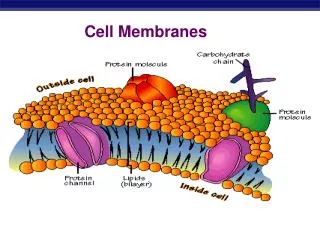
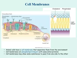
Membranes • Cells separate “inside and outside” with lipid barriers called membranes. • Organelle membranes separate too. • Limits passage of polar substances. • Protein channels allow specific passage. BILL: What is the difference between polar and charged?
What passes freely? • Small, uncharged polar molecules • Small nonpolar molecules like N2 • Model simple diffusion http://www.studyblue.com/notes/note/n/biology-172-lecture-7-flashcards/deck/124266
Cell Walls • Are outside the membrane • Structural • Plant cell walls are made of cellulose • Prokaryotes and fungi also have cell walls. http://www.phschool.com/science/biology_place/biocoach/plants/walls.html
Phospholipid Bilayer • This represents a phospholipid- • The tails are fatty acid, the head, phosphorylated alcohol • These form a sheet two molecules thick. Polar head-Hydrophilic-label Nonpolar tail-Hydrophobic-label http://en.wikipedia.org/wiki/Phospholipid
Why the embedded cholesterol? What might a membrane in an arctic dweller look like? Increase or decrease fluidity depending on temperature.(Decreases fluidity when warm, increases fluidity when cold…keeps membranes fluid at very cold temperatures)
Membrane ProteinsFunction in • Transport- • Enzyme • Surface receptors • ID Markers • Cell-cell connection • Attachment http://www.pc.maricopa.edu/Biology/pfinkenstadt/BIO201/201LessonBuilder/UnitOne/Membrane/index.html
Embedded Proteins • Can be hydrophilic with charges and polar side groups or… • Hydrophobic, with nonpolar Place your proteins in the membrane. http://www.uic.edu/classes/bios/bios100/lecturesf04am/lect08.htm
Anchoring in the membrane What could keep proteins in the membrane?
Transmembrane Proteins • Carriers- change shape • Active and passive transport • Sodium potassium pump • Channels- • are tunnels through the hydrophobic core • Receptors • Transmit information from the outside of the cell • Hormone receptors, neurotransmitters.
What needs a channel? Hydrophilic substance like large polar molecules and ions http://www.cipsm.de/en/publications/researchAreaF/2007/index.html
Carrier proteins Have you seen shape changes before? • Holds ion or molecule • Changes shape • To move something across the membrane Green ball is one K+ in a potassium pump Campbelll p 125 8th edition.
Aquaporins • Channel Protein • Each aquaporin allows 3 billion water molecules per second to pass into the cell single file. What might the positively charged region do? http://www.bio.miami.edu/~cmallery/150/memb/water.channels.htm
Receptor Proteins • Example: • G protein linked receptor • neurotransmitter
Cell Surface Markers • Glycoproteins- a carbohydrate combined with a protein. Add a carbohydrate chain to a protein embedded in the membrane. Add and label • Important in the self recognition. • Recognized by the immune system. • Glycolipid- a carbohydrate combined with a lipid . Add a carbohydrate to a lipid. Add and label. • Important in tissue recognition. • Example is blood group marker.
Cell surface markersGlycocalyx- “Sugar coating” You Me • Glycoproteins- “self” recognition The protein/carbohydrate chain shape is different person to person. For example, the major histocompatibility complex proteins are recognized by the immune system. • Glycolipids-tissue recognition The lipid/carbohydrate chain shape is specific for a certain tissue. For example blood group markers.
Transport ModesThrough the cell membrane • Passive - Down the concentration gradient-primary role in importing resources and exporting waste • Diffusion • Facilitated Diffusion- membrane proteins help charged and polar molecules pass. • Osmosis • Active- Against the concentration gradient. Energy requiring. Requires membrane proteins. • Endocytosis/Exocytosis • Na+/K+ Pump • Proton Pump
Diffusion • Often by Ion Channels • Direction of movement determined by • Relative concentration • Voltage • Each channel is specific for one or a few ions • Nervous system
Facilitated Diffusion • Carrier Proteins • Specific also • Bind/release • Moves things down the concentration gradient • Passive transport • Can become saturated
Active Transport • Uses Energy • Moves things against the gradient • Na/K Pump • Coupled Transport • Gradients created by one process can power another
Sodium Potassium Pump • Cytoplasmic Na+ binds (high affinity in this shape). • Na+ binding stimulates phosphorylation by ATP • Phosphorylation causes shape change, lower Na+ affinity, now high K+ affinity. • The K+ binding causes phosphate to be released • Phosphate release causes shape to return. Now low K+ affinity,
Endocytosis and Exocytosis • Exocytosis-internal vesicles fuse with the plasma membrane to release large macromolecules out of the cell. • Endocytosis-cell takes in macromolecules and particulate matter by forming new vesicles from the plasma membrane.
Bulk Transport • Endocytosis • Phagocytosis-particulate • Pinocytosis-liquid • Receptor-Mediated endocytosis • Clathrin coated pits bind to specific molecules.
Bulk Transport Explain the diagram. • Exocytosis • Neurotransmitter discharge • Hormone secretion • Digestion enzymes http://www.kscience.co.uk/as/module1/pictures/endoexo.jpg
Eukaryotic cells are compartmentalized By Membranes These special areas let things happen by… • Minimizing competing interactions • Increasing surface area where reactions can occur • Compartmentalizing metabolic processes and enzymatic reactions. • Examples: Endoplasmic Reticulum, mitochondria, chloroplast, Golgi, nuclear envelope • Archaea and Bacteria generally lack internal membranes and organelles.