Hydatidiform Mole
Hydatidiform Mole. Mamdoh Eskandar FRCSC. Gestational trophoblastic Disease. Molar pregnancy -Complete hydatiditform mole -Incomplete hydatiditform mole Choriocarcinoma Placental-site trophoblastic tumor. Complete mole - Fertilization an empty egg by one sperm.


Hydatidiform Mole
E N D
Presentation Transcript
Hydatidiform Mole Mamdoh Eskandar FRCSC
Gestational trophoblastic Disease • Molar pregnancy -Complete hydatiditform mole -Incomplete hydatiditform mole • Choriocarcinoma • Placental-site trophoblastic tumor
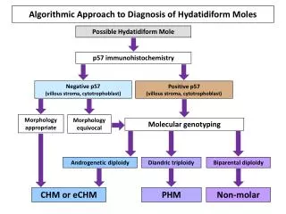
Complete mole - Fertilization an empty egg by one sperm. -All placental villa swollen. -Fetus, cord, amniotic membrane are absent. -Paternal chromosomes only. 46 XX. -diploidy Incomplete mole -fertilization of an egg by two sperms -some placental villa swollen Fetus, cord, amniotic membrane are present Paternal and maternal 69XXY -Triploid Molar Pregnancy
Molar Pregnancy • Incidence and epidemiology: -In USA 1:1000 -In Asia 8:1000 • Risk factors for molar pregnancy: -Extreme of age -Lower socioeconomic status -Race and ethnic origin -Blacks have lower incidence
Molar Pregnancy • Symptoms and signs of molar pregnancy -Abnormal bleeding in early pregnancy -Lower abdominal pain -Toxemia before 24 weeks of gestation -hyperemesis gravidarum
Molar Pregnancy -Uterus large for dates -No fetal heart rate -Enlargement of the ovaries -Hyperthyroidism -Expulsion of swollen villi
Molar Pregnancy • Diagnosis: -Ultrasound shows snowstorm-like appearance, no fetus, theca lutein cyst -Beta hCG in normal pregnancy the level is at it peak at around 14 weeks (100,000 mIU/ml)
Management • Once the diagnosis is made evacuation of the uterus should be done but prior to that: hCG preevacuation. Chest x-ray. Correct: anemia, toxemia, hyperthyroidism, pulmonary compromise.
Follow up • HCG weekly until normal for two values then monthly for one year. • Repeat x- ray if HCG rises or plateau. • Contraception for one year. • Pelvic examination every 3 weeks for 3 months.
Follow up • Initiate chemotherapy if: -HCG level is increasing or plateaus -Metastasis disease is present -HCG level is still elevated after 6 months of evacuation -HCG starts to rise after being undetectable
FICO Classification System of GTT I. Confined to corpus uteri II. Metastases to vagina or pelvic organs III. Metastases to lungs IV. Distant metastases
Prognostic Classification of GTT • I. Nonmetastatic GTT • II. Metastatic GTT: disease outside the uterus. • Good prognosis: • Disease present less than 4 months • Pretreatment HCG is less than 40,000 • No prior chemothreapy • No metastatic to the liver or the brain • Poor prognosis: the opposite of good prognosis