
Changes in FRB Domain Amide Signals Induced by Phosphatidic Acid and HTS-1 Binding
This study presents overlays of two 15N/1H HSQC spectra of the FRB domain, highlighting the shifts in backbone and side chain amide signals. The spectra were obtained from uniformly 15N-labeled FRB at a concentration of 100 µM, first measured in isolation (black) and then in the presence of saturating concentrations of phosphatidic acid (5 mM, red) and HTS-1 (0.8 mM, green). These results provide insights into the molecular interactions of the FRB domain with lipids and peptides.

Changes in FRB Domain Amide Signals Induced by Phosphatidic Acid and HTS-1 Binding
E N D
Presentation Transcript
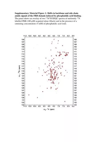
Supplementary Material Figure 1: Shifts in backbone and side chain amide signals of the FRB domain induced by phosphatidic acid binding. The panel shows an overlay of two 15N/1H HSQC spectra of uniformly 15N labelled FRB (100 µM) acquired alone (black) and in the presence of a saturating concentration (5 mM) of phosphatidic acid (red).
Supplementary Material Figure 2: Shifts in backbone and side chain amide signals of the FRB domain induced by HTS-1 binding. The panel shows an overlay of two 15N/1H HSQC spectra of uniformly 15N labelled FRB (100 µM) acquired alone (black) and in the presence of a saturating concentration (0.8 mM) of HTS-1 (green).