Download
1 / 17
240 likes | 1.4k Vues
Non invasive tissue oximetry using reflection mode Near Infrared Spectroscopy system. Dr.Ashutosh Mishra 1 and Dr.Sneh Anand 1. 1 Centre for Biomedical Engg. Periyasamy 1 Research Scholar. Indian Institute of Technology Delhi . Motivation. Oxygen –basis for human survival

E N D
Non invasive tissue oximetry using reflection mode Near Infrared Spectroscopy system Dr.Ashutosh Mishra1 and Dr.Sneh Anand1 1Centre for Biomedical Engg Periyasamy1 Research Scholar Indian Institute of Technology Delhi
Motivation • Oxygen –basis for human survival • Level of oxygen that a particular organ receives is very importance as it determines proper functioning of the body parts (organs) • For example: DM patients suffer from some form of lower extremity problem(neuropathy, vascular complication) are due to oxygen level changes(reduced blood flow) and decreased perfusion.
Importance of tissue oxygen level in any organ Deficiency of oxygen in tissue • Tissue oxygenation and hemoglobin concentration are sensitive indicators of tissue status (Ferrari, et al., 1992 ) • A sudden dip in the tissue oxygenation can be a direct indication of many harmful conditions like tissue degeneration, microbial infection etc. • Non-invasive, real time, local measurement of tissue O2 and HbT is not commercially available Tissue oxygenation - relative conc. of oxyhemoglobin & myoglobin, depends on the balance between oxygen delivery, as reflected by the product of blood flow and oxygen content and consumption.
Objectives • To design and develop an reflection mode NIRS system • To non-invasively monitor the oxygenation level in the tissue • To calculate oxy and deoxy hemoglobin concentration using Modified Beer Lambert law
Why we chosen NIR light ? Electromagnetic spectrum • Penetrate the biological tissue deeper • Property : Oxygenated hemoglobin and deoxygenated hemoglobin both absorb light differently in this region. • At 780 nm, deoxygenated blood has a higher absorption, whereas at 830 nm, oxygenated blood has a higher absorption.
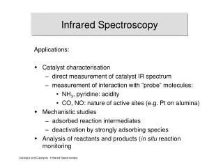
Near infrared spectroscopy Applications : • Non-invasive assessment of brain function in newborn [Wolf M et al 2007] • In-vivo muscle metabolism measurement [Yuanqing Lin et al 2002] • Monitor foetal hypoxaemia and in newborn infants to detect birth apnoea and hypoxia. • Useful for blood analyte monitoring and non invasive imaging of tissue [E. Ciurczak & J. Drennen 2002]. • Monitor healing of burns • Used for assessment and identification of breast cancer • Non-ionizing & non invasive optical technique • UV/VIS spectral region (<650), light can penetrate only superficial tissue volume [Jobsis et al 1977] • Investigates the differential absorption spectra of chromophores (oxy and deoxy hemoglobin) and functional information in tissue [Fantini et al 1999] • Measure both arterial and venous saturation because it is based on NIR wavelength(700nm-1100nm) • Healthy and diseased soft tissues can be potentially differentiated, due to their different absorption or scattering coefficients
Comparison between NIRS and Pulse oximetry • Different wavelengths are used in both these techniques • NIRS is far more penetrating effect than Pulse oximeter because sources of light is in NIR wavelength • NIRS characterize more chromophores than Pulse oximeter • NIRS - assessment of all the vascular compartments (Arterial, Venous and capillary). • Measure hemo dynamics, metabolic and fast neuronal responses to brain activation • Measure relative changes in pulsatile components of the cerebral blood flow and cerebral blood volume based on the shape of the heartbeat pulse waveform. • Used in patients with low perfusion states and peripheral vascular. It gives exact oxygen level in the blood. • Pulse oximeter - only the arterial compartment by time gating the measurements • Reliable and commonly used to monitor systemic oxygen supply only. • Pulse oximeter utilizes the arterial oscillations to extract arterial oxygen saturation SaO2 and does not exploit all of the information from the heartbeat oscillations
Design of NIR sensor Probe NIRS probe design consideration • Wavelength • Bandwidth • Power • Stability • sensitivity • Portability, ease-of-use, etc. Selection of Light source and Detector
NIR light interaction with tissue to monitor oxygenation NIR light interaction Two modes of operation Three main process: • Absorption • Scattering • Reflection. Monte Carlo Simulation of Light Transport in tissue • It is a stochastic and random process • It based on the transport equation and the random walk of photons • in absorbing and scattering medium • It provides a physical simulation of photon migration through the • tissue • Limitation • Computation time is more • Require lot of memory
Lambert Laws Lambert Beers law I = I0 10 -ε [c] L or log ( I0/I) = ε [c] L where I0 light in I light out ε extinction coefficient L the optical path length [c] solute concentration OD = - log10 (I/ I0) = ε.C.L + G Modified Beer Lambert law Where G factor that accounts for the measurement geometry L = d x B Where L optical path length value in the scattering medium. d Source-Detector separation B differential path length factor (DPF)
Methodology and Preliminary Results Feasibility study done so far… Modulating frequency waveform to drive the laser diode Probe design configuration Block diagram of NIRS system - simple prototype
Preliminary Results DATA COLLECTION : Data of two subjects(mean age 25) has been recorded with following procedure Procedure : • Step 1: The subject is first asked to relax for at least five minutes by meditation. • Step2: Then patient’s body part (forefinger) is cleaned to avoid any unnecessary disturbance by dirt etc in the light reflection process. • Step 3: Now we place the forefinger on the NIRS probe as shown in fig “with out occlusion” and switch on the power supply to observe the change in light intensity in the Oscilloscope (CRO). • Step 4:Then note down the peak voltage (in mV) value from the CRO. • Step 5: Once the occlusion is done for 5-10 seconds by using BP cuff, then proceed the step 3 and 4 respectively Placement Fore finger over NIRS probe
Result Calculation Peak value (mV) “With” and “Without occlusion” Calculation of Change in optical density Optical path length L =0.6 cm Δ [OHb] = [(εDHb830X ΔOD780) - (ε DHb780 X ΔOD830)] / [L ( εOHb780 X εDHb830 - εOHb830 X εDHb780)] Δ [OHb] = 83.6 µM/L ε Extinction coefficient taken from literature Method of Calculation Δ [DHb] = [(εOHb780X ΔOD830) - (ε OHb 830X ΔOD780)] / [L ( εOHb780 X εDHb830 – εOHb830 X εDHb780)] Δ [DHb] =10.5 µM/L Δ [THb] = Δ [OHb] + Δ [DHb] Δ [THb] =83.6+10.5=94.1µM/L Tissue oxygenation Index TOI = (Δ [OHb] / Δ [THb]) x100 TOI = 88.8%
References • [1] J.Mobley and T.Vo-Dinh, Optical properties of tissue, Biomedical photonics handbook, CRC Press 2005, pp. 2-1-2-74 • [2] M. Cutler, Transillumination of the breast, Surg. Gynecol. Obstet. Vol. 48, 1929, 721-727. • [3] Liss, J.M., White, L., Mattys, S., Spitzer, S., Lansford, K., Lotto,A.J., and Caviness, J.Classifying Dysarthrias by Speech Rhythm Metrics. Auditory Cognitive, Neuroscience Society (ACNS) Conference 2009 • [4] Alper Bozkurt, Arye Rosen, Hare Rosen and Banu Onaral A portable near infrared spectroscopy system for bedside monitoring of newborn brain, BioMedical Engineering OnLine 2005, 4:29 • [5] A.Pifferi, P.Taroni, G.Valentini and S.Andersson-Engels, Real-time method for fitting time-resolved reflectance and transmittance measurements with a Monte Carlo model, Appl.Opt 1998, 37, 2774-2780. • [6] Jöbsis F.F, Noninvasive infrared monitoring of cerebral and myocardial oxygen sufficiency and circulatory parameters, Science 1977,198: pp. 1264–1267 • [7] E. Ciurczak and J. Drennen, Near-Infrared Spectroscopy in Pharmaceutical and Medical Applications, Marcel-Dekker, Inc. New York, 2002. • [8] Wolf M, et al. Progress of near infrared spectroscopy and imaging instrumentation for brain and muscle clinical applications. J. Biomed. Opt. 2007; 12, 062104. Review
Thank You Centre for Biomedical Engg Indian Institute of Technology Delhi