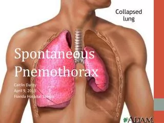
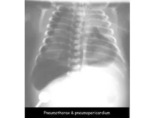
Pneumothorax & pneumopericardium
Pneumothorax & pneumopericardium. Pneumothorax, pneumopericardium pneumoperitoneum, subcut. emphysema. Pulmonary interstitial emphysema. CATHETER AND TUBE POSITION. 1. ENDOTRACHEAL TUBE : 1 cm below vocal cord & 2 cm above carina 2. NASOGASTRIC TUBE : In stomach

Pneumothorax & pneumopericardium
E N D
Presentation Transcript
Pneumothorax, pneumopericardium pneumoperitoneum, subcut. emphysema
CATHETERANDTUBEPOSITION 1. ENDOTRACHEAL TUBE : 1 cm below vocal cord & 2 cm above carina 2. NASOGASTRIC TUBE : In stomach 3. UVC : in IVC or RA proper 4. UAC : in thoracic aorta below level of DA ( T4 ) and above origin of celiac artery ( T11 )
RESPIRATORY DISTRESS IN NEWBORNSURGICAL CAUSES DIAPHRAGMATIC HERNIA • Common on left side • Multiple lucencies or cysts ( bowel loops ) in the chest • Shifting of heart and mediastinum to the other side • Decreased bowel gas or gasless abdomen
FOREIGN BODY ASPIRATION • Common in children 1 - 3 years old • Tracheal obstruction • Bronchial obstruction Incomplete - check valve : Air trapping Complete : Atelectasis • S/S : Dyspnea, hyperpnea, cough, +/- Hx of FB aspiration
F 11 mo , หายใจหอบหลังกินถั่วติดคอมา 1อาทิตย์
Scope : peanut in Rt.main bronchus Post F.B. removal film
F 4mo, cough, หายใจหอบหลังสำลักเม็ดส้มมา 1 wk
ALIMENTARY TRACT OBSTRUCTION IN NEWBORN Normal abdominal bowel gas pattern : ‘Polygonal pattern’ • Stomach : immediate after birth • Small bowel : within 3 hours • Colon : within 5 hours • Rectum : within 6-8 hours • Pronefilm to demonstrate air in rectum ( R/O obstruction ) • Upright or left lateral decubitus film to demonstrate free air
ESOPHAGEAL ATRESIA ( EA ) • MC = proximal eso. atresia with distal TEF • High incidence of associated anomalies : VACTERL ( V-vertebral, A-anorectal , C-cardiovascular , TE-tracheoesophageal ,R-renal , L-limb )
GASTRIC OUTLET OBSTRUCTIONIDIOPATHIC HYPERTROPHIC PYLORIC STENOSIS S/S : Non-billous vomitting Onset ~ 1 month old Minimal upper abdominal distension +/-palpable mass ( olive ) at epigastrium
IDIOPATHIC HYPERTROPHIC PYLORIC STENOSIS ( IHPS ) X-RAY FINDINGS : • Plain film : Gastric dilatation with large amount of air/fluid content , Decreased small bowel gas • US : Elongation with thickening of muscle wall of pyloric canal • UGI : Narrowing and elongation of pyloric canal ‘String sign’ Muscle indentation on lumen of stomach and duodenum ‘shoulder sign’