Download

1 / 39
420 likes | 664 Vues
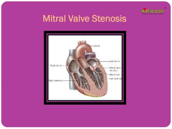
HAEMODYNAMICS OF MITRAL STENOSIS. DR VINOD G V. Normal MVA 4-5 cm2 No pressure gradient across mitral valve during diastole Consequence of narrowed orifice 1.Development of pressure gradient across mitral valve 2.Progressive rise in LA pressure, pulmonary venous pressure

E N D
HAEMODYNAMICS OF MITRAL STENOSIS DR VINOD G V
Normal MVA 4-5 cm2 • No pressure gradient across mitral valve during diastole Consequence of narrowed orifice 1.Development of pressure gradient across mitral valve 2.Progressive rise in LA pressure, pulmonary venous pressure 3.Dependence of LV filling on LA pressure 4.Reduction of blood flow across mitral valve
Torricelli's 2 Torricelli's 1 F=CO/HR xDFP
Factors ↑ gradient • ↑ COP • Exertion ,emotion,high output states • ↓ DFP • Increase HR • ↓ MVA • Progression of disease • Factors decreasing gradient • ↓ COP • Second stenosis • RV failure • ↑ DFP • Slow HR • ↑ MVA
PAH in MS • Passive -Obligatory increase in response to increased LA pressure to maintain gradient of 10 to 12 across pulmonary vascular bed(PA mean- LA mean) • Reactive PA mean pressure –LA mean pressure >10 to 12 Pulmonary vasoconstriction • Obliterative changes in pulmonary arterioles Medial hypertrophy Intimal proliferation
Causes of reactive pulmonary HTN • Wood-pulmonary vasoconstriction • Doyle-↑pulmonary venous pressure prominent in the lower lobes, produce reflex arterial constriction • Heath &Harris-↑ PA pressure causes reflex arteriolar constriction
Jordan- • ↑pulmonary venous pressure-transudation of fluid • causes thickening and fibrosis of alveolar walls • hypoventilation of lower lobes-hypoxemia in lower lobe vessels • Sensed by chemoreceptors in pulmonary veins • Pulmonary arteriolar vasoconstriction in regions supplying these alveoli • Lower lobe perfusion decreases • This process eventually involve middle and upper lobe
Stage 1 • Asymptomatic at rest • Stage 2 • Symptomatic due to elevated LA pressure • Normal pulmonary vascular resistance • Stage 3 • Increased pulmonary vascular resistance • symptoms of low COP • Stage 4 • Both stenosis severe • Extreme elevation of PVR-RV failure
Consequence of PAH: • RVH,TR • Reduced CO • Elevated pre capillary resistance protects against development of pulmonary congestion at cost of a reduced CO • Severe pulmonary HTN leads to right sided failure
Effect of AF • ↑HR,↓DFP-elevates trans mitral gradient • Can result in acute pulmonary edema • Loss of atrial contribution to LV filling • Normal contribution of LA contraction to LV filling 15% • In MS, increases up to 25-30% • Lost in AF
Calculation of MVA Gorlin’s formula
Flow • Total cardiac output divided by time in seconds during which flow occurs across the valve • F=COP/DFPXHR
Steps in calculating MVA • Average gradient=area(mm2)/length of diastole(mm) • Mean gradient=average gradient X scale factor • Average diastolic period=length of DFP(mm)/paper speed(mm/s) • HR(beat/min), COP(ml/min) • MVA=cardiac output/HR × average diastolic periodperiod÷37.7×√mean gradient
Pitfalls in calculating MVA • Overestimation of trans mitral gradient occurs when PCWP is not taken properly • Failure to wedge properly cause one to compare damped pulmonary artery pressure to LV pressure • To ensure proper wedging -mean wedge pressure is lower than mean PA pressure -Blood withdrawn from wedge catheter is >95% saturated
Alignment Mismatch • Alignment of the PCW and LV pressure tracings does not match alignment of simultaneous LA and LV tracings • There is a time delay of 50-70msec • V wave in LA pressure tracing peaks immediately before LV pressure down stroke • Realign wedge tracing so that the V wave peak is bisected by or slightly to the left of the down stroke of LV pressure
CO determination • Simultaneous measurement with LA-LV pressure tracing • Under estimation of valve area in case of associated MR • Thermo dilution method inaccurate when associated TR
Damped PCW-LV Vs LA-LV Overestimation of MVG occur if damped PCW P is used
LA-LV gradient in AF • With long diastolic filling period ,progressive decrease in LA pressure • Increase with short diastole • Measure gradient in 3 to 4 diastolic complexes with nearly equal cycle length and measure the mean value
Symptoms and signs Hemodynamic correlation
Acute pulmonary edema • Increased pulmonary venous pressure • Increased transudation of fluid • Decreased lymphatic clearance • Pulmonary capillary pressure exceeds tissue oncotic pressure of 25mm Hg
Hemoptysis • Pulmonary apoplexy -rapture of bronchial vein -massive hemoptysis • Pink frothy sputum during pulmonary edema • Chronic bronchitis • Pulmonary infarction
Loud S1 • Rapidity with which LV pressure rises when mitral valve closes • Mitral valve closes at higher dp/dt of LV • Wide closing excursion of valve leaflets
A2-OS interval • OS occurs due to sudden tensing of valve leaflets after the valve cusps have completed their opening excursion • Follows A2 by 40-120msec • Interval varies inversely with LA pressure • Shorter A2-OS interval indicates severe MS
Diastolic murmur • Mid diastolic component starts with OS • Holo diastolic in severe MS due to persistent gradient • Presystolic component: -Atrial contraction -Persistent LA-LV pressure gradient - can persists even in AF
Doppler ECHO • Rate of fall in flow velocity is slow • No period of diastasis • Increased early diastolic peak velocity
Mitral Pressure Half Time • The pressure halftime is defined as the time required for the pressure to decay to half its original value • Mitral valve area (MVA) calculated as: MVA = 220/PHT • Not affected by CO,MR • PHT =11 .6xCnx√ MPG/(CcxMVA) • Cn-net compliance
Disadvantage • Poor ventricular compliance will increase the rate of pressure rise in diastole • Shorten PHT overestimate MVA • Significant AR, diastolic dysfunction alter PHT • Post BMV PHT is inaccurate
Q1 • O2 consumption 180 ml/min • A-V O2 difference 40 ml/L • HR 76/min SR • LV diastolic mean 6 • Diastolic filling period 0.42 sec/beat • PCW mean 24 • PA 40/22 -mean 22
Q2 • Body surface area 1.4 m2 • O2 consumption 201ml/min • A-V O2 difference 110 mL/L • PR 92/min • LV diastolic mean 10 • Diastolic filling period 0.36sec/beat • PCW mean 33 • PA 125/65, mean 75