Download
1 / 74
970 likes | 1.61k Vues
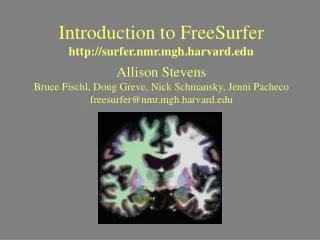
Introduction to FreeSurfer http://surfer.nmr.mgh.harvard.edu. Allison Stevens Bruce Fischl, Doug Greve, Nick Schmansky, Jenni Pacheco freesurfer@nmr.mgh.harvard.edu. Cortical Surface Reconstruction. FreeSurfer creates computerized models of the brain from MRI data. Input:

E N D
Introduction to FreeSurferhttp://surfer.nmr.mgh.harvard.edu Allison Stevens Bruce Fischl, Doug Greve, Nick Schmansky, Jenni Pacheco freesurfer@nmr.mgh.harvard.edu
Cortical SurfaceReconstruction FreeSurfer creates computerized models of the brain from MRI data. Input: • T1-weighted (MPRAGE,SPGR) • 1mm3 resolution
Cortical SurfaceReconstruction • Finds white/gray boundary – wm surface • Finds pial/CSF boundary – pial surface • To “Find” uses: • Intensity information, spatial location, geometric structure • Tessellation, neighbors, talairach coordinates • Subcortical Segmentation
Surface Model • Mesh (“Finite Element”) • Vertex = point of 6 triangles • XYZ at each vertex • Triangles/Faces ~ 150,000 • Area, Distance • Curvature, Thickness
Cortical Reconstruction Goals • Geometrically Accurate surfaces • Accurately follow the boundaries seen on the scan for each of your individual subjects • Topologically Correct surfaces • Each surface is a 2-D continuous, non self-intersecting sheet and can be inflated into a perfect sphere • Surfaces are only as good as your scan.
MR Anatomy Caveats • Dependent on data quality • Contrast to noise • Signal to noise • Voxel resolution • MR Artifacts • MR susceptibility • MR distortions • Variations in MR tissue parameters across regions of the brain are altered in different populations
FreeSurfer Output • Volumes • Surfaces • Surface Overlays • ROI Summaries
Volumes orig.mgz T1.mgz brainmask.mgz wm.mgz filled.mgz Subcortical Mass • $SUBJECTS_DIR/bert/mri • All “Conformed” 256^3, 1mm aseg.mgz aparc+aseg.mgz Volume Viewer: tkmedit
orig white pial Surfaces inflated sphere,sphere.reg flat • $SUBJECTS_DIR/bert/surf • Number/Identity of vertices stays the same (except flat) • XYZ Location Changes • Flattening not done as part of standard reconstruction Surface Viewer: tksurfer
Surface Overlays lh.thickness on inflated lh.sulc on inflated lh.curv on inflated lh.curv on inflated lh.sulc on pial fMRI on flat • Value for each vertex • Color indicates value • Color: gray,red/green, heat, color table • Rendered on any surface • fMRI/Stat Maps too lh.aparc.annot on inflated
ROI Summaries • aseg.stats • volumes of subcortical structures (mm3) • aparc.stats • thickness of cortical parcellation structures (mm) • total white matter volume (mm3) • number of vertices in cortex • surface area of cortex (mm2)
ROI Summaries: Make Your Own yourROI.label • Draw your own surface label • use mris_anatomical_stats to get data mris_volume • Total volume within a surface you specify mris_wm_volume • Total volume within white surface ignoring non-wm voxels in aseg.mgz
Reconstruction Environment • Installation directory: $FREESURFER_HOME • Set-up Environmental Variables • Unix command-line (Linux, MacOSX) • Directory structure, naming conventions • File Formats
Set-up Environmental Variables Subject ID $SUBJECTS_DIR bert fred sara margaret … Before running FreeSurfer, must set $FREESURFER_HOME and $SUBJECTS_DIR
FreeSurfer Directory Tree Each data set has its own unique SubjectId (eg, bert) Subject ID bert bem stats src mri scripts surf tmp label trash
FreeSurfer Directory Tree Directories used often are in green. Subject ID bert bemstatssrcmriscriptssurftmplabel trash aseg.stats lh.aparc.stats rh.aparc.stats wmparc.stats
MGZFile Format 001.mgz • mgz = compressed MGH file • Can store 4D (like NIFTI) • cols, rows, slices, frames • Generic: volumes and surfaces • Eg, Typical Anatomical volume: 256 x 256 x 128 x 1 lh.thickness.sm10.mgz • FreeSurfer can read: • DICOM, Siemens IMA, AFNI • FreeSurfer can read/write: • NIFTI, Analyze, MINC Careful with NIFTI! (32k limit)
Other FreeSurfer File Formats • Unique to FreeSurfer • Surface: lh.white, lh.pial, lh.orig • Curv: lh.curv, lh.sulc, lh.thickness • Annotation: lh.aparc.annot • Label: lh.pericalcarine.label
Starting the Reconstruction Process Before running FreeSurfer, must set $FREESURFER_HOME and $SUBJECTS_DIR recon-all -i /path/to/your/raw/data1 -i /path/to/your/raw/data2 -all-s subject_id • This will create the subject directory ‘subject_id’ in your $SUBJECTS_DIR and convert your 2 raw acquisitions to mgz and use them as input for the ‘-all’ command.
Alternative: AddYourData • cd $SUBJECTS_DIR • mkdir –p bert/mri/orig • mri_convert yourdicom.dcm bert/mri/orig/001.mgz • mri_convert yourdicom.dcm bert/mri/orig/002.mgz • recon-all –all –s bert bert bemlabelsrcmriscriptssurf tmplabel orig 001.mgz002.mgz
Individual Steps Volumetric Processing Stages (subjid/mri): 1. Motion Cor, Avg, Conform (orig.mgz) 2. Non-uniform inorm (nu.mgz) 3. Talairach transform computation (talairach/talairach.xfm) 4. Intensity Normalization 1 (T1.mgz) 5. Skull Strip (brainmask.mgz) 6. EM Register (linear volumetric registration) 7. CA Intensity Normalization (norm.mgz) 8. CA Non-linear Volumetric Registration 9. CA Label (Volumetric Labeling) (aseg.mgz) 10. Intensity Normalization 2 (T1.mgz) 11. White matter segmentation (wm.mgz) 12. Edit WM With ASeg 13. Fill and cut (filled.mgz) Surface Processing Stages (subjid/surf): 14. Tessellate (?h.orig.nofix) 15. Smooth1 16. Inflate1 17. QSphere (?h.qsqhere) 18. Automatic Topology Fixer (?h.orig) 19. Final Surfs (?h.white ?h.pial ?.thickness) 20. Smooth2 (?h.smoothwm) 21. Inflate2 (?h.inflated) 22. Aseg Statistics (stats/aseg.stats) 23. Cortical Ribbon Mask (?h.ribbon.mgz) 24. Spherical Morph 25. Spherical Registration (?h.sphere.reg) 26. Map average curvature to subject 27. Cortical Parcellation (Labeling) 28. Cortical Parcellation Statistics 29. Cortical Parcellation mapped to Aseg 30. White Matter Parcellation (wmparc.mgz) Blue = Manual Intervention recon-all -help Note: ?h.orig means lh.orig or rh.orig
Reconstrution Stages recon-all is broken into three stages • autorecon1 • autorecon2 • autorecon3
-autorecon1 Volumetric Processing Stages (subjid/mri): 1. Motion Cor, Avg, Conform (orig.mgz) 2. Non-uniform inorm (nu.mgz) 3. Talairach transform computation (talairach/talairach.xfm) 4. Intensity Normalization 1 (T1.mgz) 5. Skull Strip (brainmask.mgz) 6. EM Register (linear volumetric registration) 7. CA Intensity Normalization (norm.mgz) 8. CA Non-linear Volumetric Registration 9. CA Label (Volumetric Labeling) (aseg.mgz) 10. Intensity Normalization 2 (T1.mgz) 11. White matter segmentation (wm.mgz) 12. Edit WM With ASeg 13. Fill and cut (filled.mgz) Surface Processing Stages (subjid/surf): 14. Tessellate (?h.orig.nofix) 15. Smooth1 16. Inflate1 17. QSphere (?h.qsqhere) 18. Automatic Topology Fixer (?h.orig) 19. Final Surfs (?h.white ?h.pial ?.thickness) 20. Smooth2 (?h.smoothwm) 21. Inflate2 (?h.inflated) 22. Aseg Statistics (stats/aseg.stats) 23. Cortical Ribbon Mask (?h.ribbon.mgz) 24. Spherical Morph 25. Spherical Registration (?h.sphere.reg) 26. Map average curvature to subject 27. Cortical Parcellation (Labeling) 28. Cortical Parcellation Statistics 29. Cortical Parcellation mapped to Aseg 30. White Matter Parcellation (wmparc.mgz) recon-all -help
-motioncor -autorecon1 mri orig rawavg.mgz 001.mgz 002.mgz Motion Correction and Averaging 001.mgz rawavg.mgz + 002.mgz Does not change native resolution. mri_motion_correct.fsl
-motioncor -autorecon1 rawavg.mgz orig.mgz Conform Changesto 256^3, 1mm^3 All volumes will be conformed. orig Volume mri_convert -conform
-nuintensitycor -autorecon1 Non-Uniform Intensity Correction • Uses MNI tool • Removes B1 bias field nu Volume mri_nu_correct.mni
-talairach -autorecon1 Talairach Transform • Computes 12 DOF transform matrix • Does NOT resample • MNI305 template • Used to help find structures (eg, CC) • Can also be used to localize functional activation • mri/transforms/talairach.xfm mri transforms talairach.xfm talairach_avi
-normalization -autorecon1 IntensityNormalization • Presegmentation (T1.mgz) • All WM = 110 intensity • Pre- and Post-Skull Strip T1 Volume mri_normalize
-skullstrip -autorecon1 SkullStrip • Removes all non-brain • Skull, Eyes, Neck, Dura • brainmask.mgz Orig Volume Brainmask Volume mri_watershed
FreeSurfer Directory Tree Each data set has its own unique SubjectId (eg, bert) Subject ID bert bem stats srcmriscripts surf tmp label trash orig T1 brainmaskwmaseg aparc+aseg wmparc
-autorecon2 Volumetric Processing Stages (subjid/mri): 1. Motion Cor, Avg, Conform (orig.mgz) 2. Non-uniform inorm (nu.mgz) 3. Talairach transform computation (talairach/talairach.xfm) 4. Intensity Normalization 1 (T1.mgz) 5. Skull Strip (brainmask.mgz) 6. EM Register (linear volumetric registration) 7. CA Intensity Normalization (norm.mgz) 8. CA Non-linear Volumetric Registration 9. CA Label (Volumetric Labeling) (aseg.mgz) 10. Intensity Normalization 2 (T1.mgz) 11. White matter segmentation (wm.mgz) 12. Edit WM With ASeg 13. Fill and cut (filled.mgz) Surface Processing Stages (subjid/surf): 14. Tessellate (?h.orig.nofix) 15. Smooth1 16. Inflate1 17. QSphere (?h.qsqhere) 18. Automatic Topology Fixer (?h.orig) 19. Final Surfs (?h.white ?h.pial ?.thickness) 20. Smooth2 (?h.smoothwm) 21. Inflate2 (?h.inflated) 22. Aseg Statistics (stats/aseg.stats) 23. Cortical Ribbon Mask (?h.ribbon.mgz) 24. Spherical Morph 25. Spherical Registration (?h.sphere.reg) 26. Map average curvature to subject 27. Cortical Parcellation (Labeling) 28. Cortical Parcellation Statistics 29. Cortical Parcellation mapped to Aseg 30. White Matter Parcellation (wmparc.mgz) recon-all -help Note: lh processed completely first, then rh.
-subcortseg -autorecon2 Automatic Volume Labeling • Used to determine volumes of subcortical structures • Used to fill in subcortical structures for creating subcortical mass • aseg.mgz ASeg Volume Atlas: RB_all_2007-08-08 steps 6-9, 22
-subcortseg -autorecon2 Volume-based Labeling Labeling isdetermined by location and intensity.
Validation of Volume Labeling * Manual labeling done by Center for Morphometric Analysis (CMA) *Thanks to Drs Larry Seidman and Jill Goldstein for providing this data.
Find “Subcortical Mass” • All White Matter • All Subcortical Structures • Ventricles • Excludes brain stem and cerebellum • Hemispheres separated • Completely connected (no islands) • Many Stages … More Later …
-segmentation -autorecon2 wm Volume White Matter Segmentation • Separates white matter from everything else • “Fills in” subcortical structures • Cerebellum removed, brain stem still there mri_segment mri_edit_wm_with_aseg mri_pretess
-fill -autorecon2 WM Volume Filled Volume Fill and Cut (Subcortical Mass) • Fills in any voids • Removes any islands • Removes brain stem • Separates hemispheres (each hemi has different value) • filled.mgz = “Subcortical Mass” mri_fill
FreeSurfer Directory Tree Each data set has its own unique SubjectId (eg, bert) Subject ID bert bem stats src mriscripts surf tmp label trash orig T1 brainmask wm aseg aparc+aseg wmparc
Tessellation -tessellation -autorecon2 orig surface surf/lh.orig surf/rh.orig • Mosaic of triangles (“tessellation”) • Errors: Donut holes, handles • - Subsequently fixed by the automatic topology fixer
-inflate -autorecon2 Inflation: Visualization Gyri Sulci Dale and Sereno, 1993; Dale et al., Dale et al., 1999; Fischl et al., 1999; Fischl et al., 2000; Fischl et al., 2001
Automatic Topology Fixer -fix -autorecon2 Fornix hippocampus optic nerve Ventricles and Caudate Cortical Defects Pallidum and Putamen • Holes • Handles • Automatically Fixed
White Matter Surface -finalsurfs -autorecon2 • Nudge orig surface • Follow T1 intensity gradients • Smoothness constraint • Vertex Identity stays constant
Pial Surface -finalsurfs -autorecon2 • Nudge white surface • Follow T1 intensity gradients • Vertex Identity Stays
-finalsurfs -autorecon2 Optimal Surface Placement Gray-WhiteBoundary OuterCorticalSurface
-autorecon2 Gray/CSF Deformation Gray-WhiteBoundary OuterCorticalSurface Dale and Sereno, 1993; Dale et al., Dale et al., 1999; Fischl et al., 1999; Fischl et al., 2000; Fischl et al., 2001
-finalsurfs -autorecon2 Cortical Thickness
Thickness Maps • Red regions are thinner • Yellow regions are thicker
4 3.5 3.66 3.61 3.22 3.27 3.45 3.17 3.37 3.04 3.5 3.16 3.16 3.14 3 3.03 3 2.62 3 2.6 2.57 2.73 2.52 2.53 2.48 2.44 2.42 2.41 2.5 2.36 2.44 2.39 2.5 2.23 2.14 2 2 1.5 1.5 1 1 0.5 0.5 0 0 Histological Validation 4.0 0 3.5 0 Cortical Thickness (mm) Cortical Thickness (mm) Cortical Thickness (mm) Cortical Thickness (mm) Cun Fus IP IF PC MF ST PrC Cun IP SM MF PST IF Brain Region (HD) Brain Region Brain Region Brain Region (Normal) Courtesy of Diana Rosas, MGH (Rosas, et al., 2002, Neurology)
FreeSurfer Directory Tree Each data set has its own unique SubjectId (eg, bert) Subject ID bert bem stats src mri scripts surf tmp label lh.white lh.curvlh.thicknesslh.pial
-autorecon3 Volumetric Processing Stages (subjid/mri): 1. Motion Cor, Avg, Conform (orig.mgz) 2. Non-uniform inorm (nu.mgz) 3. Talairach transform computation (talairach/talairach.xfm) 4. Intensity Normalization 1 (T1.mgz) 5. Skull Strip (brainmask.mgz) 6. EM Register (linear volumetric registration) 7. CA Intensity Normalization (norm.mgz) 8. CA Non-linear Volumetric Registration 9. CA Label (Volumetric Labeling) (aseg.mgz) 10. Intensity Normalization 2 (T1.mgz) 11. White matter segmentation (wm.mgz) 12. Edit WM With ASeg 13. Fill and cut (filled.mgz) Surface Processing Stages (subjid/surf): 14. Tessellate (?h.orig.nofix) 15. Smooth1 16. Inflate1 17. QSphere (?h.qsqhere) 18. Automatic Topology Fixer (?h.orig) 19. Final Surfs (?h.white ?h.pial ?.thickness) 20. Smooth2 (?h.smoothwm) 21. Inflate2 (?h.inflated) 22. Aseg Statistics (stats/aseg.stats) 23. Cortical Ribbon Mask (?h.ribbon.mgz) 24. Spherical Morph 25. Spherical Registration (?h.sphere.reg) 26. Map average curvature to subject 27. Cortical Parcellation (Labeling) 28. Cortical Parcellation Statistics 29. Cortical Parcellation mapped to Aseg 30. White Matter Parcellation (wmparc.mgz) recon-all -help Note: lh processed completely first, then rh.