Download
1 / 65
650 likes | 819 Vues
Clinical Pathology Conference “62 year old woman with weakness and shortness of breath”. Heather Henderson, MD Internal Medicine Resident, PGY-3 Scott & White/Texas A&M HSC. Case Presentation. CC: “I am so weak and short of breath” HPI: 62 year old white woman with multiple sclerosis

E N D
Clinical Pathology Conference“62 year old woman with weakness and shortness of breath” Heather Henderson, MD Internal Medicine Resident, PGY-3 Scott & White/Texas A&M HSC
Case Presentation • CC: “I am so weak and short of breath” • HPI: 62 year old white woman with multiple sclerosis • 2-3 days of worsening lower extremity swelling • Increasing shortness of breath • Generalized weakness • No prior history of heart failure symptoms
Case Presentation • Past Medical History: • Relapsing, remitting multiple sclerosis • HTN • History of herpes zoster • Raynaud’s phenomenon • S/P TAH/BSO
Case Presentation • Allergies: NKDA • Medications: • Amantadine 200 mg po qam • Avonex 30 mcg IM qweekly • Baclofen 10 mg po qid • Amitriptyline 50 mg po qday • Oxybutynin 5 mg po bid • Maxzide 75/50 po daily, but has not taken for last week • Conjugated estrogen 0.625 mg po daily
Case Presentation • Social History: • Tobacco: None • ETOH: None • Lives alone in Temple • Inactive, but performs ADL’s • Family History: • Prostate Ca – brother • HTN - brother
Case Presentation • Review of Systems: • Weak with poor appetite • Dyspnea on exertion with some wheezing lately • No fever or chills • Constipated for last 2-3 days • Nauseated at times without emesis • No headaches or blurred vision • No dysuria • No recent problem with Raynaud’s
Physical Examination • VS:151/60, 120, 20, 93% RA, 36.1, Wt: 91kg • Gen: wn/wd white woman appearing chronically ill, weak, and tired, but no acute distress • HEENT: normal except JVP =10 cm of water • Chest: bilateral breath sounds decreased bilateral lower lobes, bibasilar crackles • CV: PMI slightly displaced inferolaterally; tachycardic at 120 bpm; normal S1 and S2 with normal splitting, no S3 or S4 gallop; II/VI diastolic decrescendo murmur at LLSB with patient sitting up; pulses 2+/2+, bilateral throughout
Physical Examination • Back: pitting sacral edema • Abd: soft, nd/nt; nabs, no abd bruit, no hepatosplenomegaly • Ext: no clubbing/cyanosis; positive 0.5cm depth pitting edema of the lower extremities L>R to the level of the upper tibia • Skin: chronic venous stasis changes bilateral lower extremities • Neuro: bulk and tone normal in UE’s, LE’s with decrease motor strength with patient unable to lift left leg (known to be chronic); no new sensory deficits
Laboratory Evaluation • BNP=1940 • TnI=0.07, CK=35, CK-MB=1.8 • Na=140, K=3.7, Cr=1.0, BUN=6 • TSH=1.3 • WBC=12.7, Hbg=13.9, Plat=391k • Chol=178, TG=152, HDL=67, LDL=81
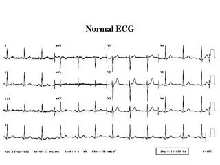
Electrocardiogram • NSR, rate = 84 • Marked t-wave inversion in the anterior leads and scooping of the ST segment in the inferior leads, suggests ischemia • No voltage criteria for LVH
Chest X-ray • Cardiomegaly • Pulmonary vascular congestion • Small bilateral pleural effusions
Echocardiogram • LV enlargement with EF=25% (globally depressed) • Normal LV wall thickness • Increased echo densities in the LV apex suggesting possible thrombus • LA enlargement with mild MR • Moderate to severe AI with normal aortic root size
Transesophageal Echocardiogram • AI was mild to moderate • Prominent myocardial trabeculations • No LV thrombus
Coronary Angiogram • Normal coronary anatomy • No significant angiographic coronary artery disease
Weakness Shortness of breath Multiple Sclerosis Congestive Heart Failure Dilated Cardiomyopathy EF =25% Tachycardia Aortic Insufficiency with normal aortic root Prominent myocardial trabeculations No significant Coronary Artery Disease Problem List
Objectives • Define cardiomyopathy • Discussion of causes of dilated cardiomyopathy • Diagnostic evaluation of a dilated cardiomyopathy • Review of the literature for a correlation between multiple sclerosis and cardiomyopathy • A discussion of a rare cause of dilated cardiomyopathy
Cardiomyopathy Defined • A group of disorders in which the dominant feature is direct involvement of the heart muscle. • (Not the result of pericardial, hypertensive, congenital, or valvular diseases)
Classification of Primary Cardiomyopathies • Dilated cardiomyopathy • Hypertrophic cardiomyopathy • Restrictive cardiomyopathy • Arrhythmogenic right ventricular cardiomyopathy • Unclassified cardiomyopathy
Specific Cardiomyopathies • Ischemic cardiomyopathy • Valvular cardiomyopathy • Hypertensive cardiomyopathy • Inflammatory cardiomyopathy • Metabolic cardiomyopathy • General-systemic disease cardiomyopathy • Muscular dystrophies • Neuromuscular disorders • Sensitivity and toxic reactions • Peripartal cardiomyopathy
Dilated Cardiomyopathy • 5-8 cases per 100,000 population/year • 10,000 deaths each year in the US • 46,000 hospitalizations each year in the United States • ¼ of the cases of congestive heart failure in the United States • 75 different diseases cause DCM
Objectives • Define cardiomyopathy • Discussion of causes of dilated cardiomyopathy • Diagnostic evaluation of a dilated cardiomyopathy • Review of the literature for a correlation between multiple sclerosis and cardiomyopathy • A discussion of a rare cause of dilated cardiomyopathy
Ischemia Infectious diseases Coxsackievirus Cytomegalovirus HIV Varicella Hepatitis Epstein-Barr Echovirus Streptococci-rheumatic fever Typhoid fever Diphtheria Brucellosis Psittacosis Rickettsial disease Lyme disease Histoplasmosis Cryptococcosis Toxoplasmosis Trypanosomiasis Shistosomiasis Trichinosis Causes of Dilated Cardiomyopathy
Medications Chemotherapeutic agent Anthracyclines Cyclophosphamide Trastuzumab Antiretroviral drugs Zidovudine Didanosine Zalcitabine Phenothiazines Chloroquine Clozapine Toxins Ethanol Cocaine Amphetamines Cobalt Lead Mercury Carbon Monoxide Beryllium Causes of Dilated Cardiomyopathy
Rheumatologic diseases Systemic lupus Scleroderma Giant cell arteritis Endocrinologic disorders Hypo/Hyperthyroidism Growth hormone excess or deficiency Pheochromocytoma Diabetes Mellitus Cushing’s disease Neuromuscular diseases Duchenne’s Muscular Dystrophy Myotonic dystrophy Friedreich’s ataxia Deposition Disease Hemochromatosis Amyloidosis Causes of Dilated Cardiomyopathy
Electrolyte abnormalities Hypocalcemia Hypophosphatemia Uremia Nutritional deficiencies Thiamine Selenium Carnitine Miscellaneous Peripartum cardiomyopathy Tachycardia Sarcoidosis Familial Sleep Apnea Autoimmune myocarditis Radiation Calcium Overload Oxygen free radical damage Causes of Dilated Cardiomyopathy
Frequency of Different Causes • Idiopathic – 50 percent • Myocarditis – 9 percent • Ischemic heart disease – 7 percent • Infiltrative disease – 5 percent • Peripartum cardiomyopathy – 4 percent • HIV infection – 4 percent • Connective tissue disease – 3 percent • Substance abuse – 3 percent • Doxorubicin – 1 percent • Other – 10 percent
Differential Diagnosis • Dilated Cardiomyopathy • Idiopathic dilated cardiomyopathy • Valvular cardiomyopathy • Medications • Multiple Sclerosis • Left ventricular noncompaction
Objectives • Define cardiomyopathy • Discussion of causes of dilated cardiomyopathy • Diagnostic evaluation of a dilated cardiomyopathy • Review of the literature for a correlation between multiple sclerosis and cardiomyopathy • A discussion of a rare cause of dilated cardiomyopathy
Noninvasive Laboratory Evaluation • Ca • Phos • Creatinine, BUN • Thyroid function studies • Iron studies • HIV
Invasive Evaluation • Endomyocardial biopsy • May be of benefit in certain situations • Definite clinical benefit • Infiltrative disorders • Anthracycline toxicity • Cardiac transplant rejection • No definitive pattern histologically in DCM • Estimated that a specific diagnosis is obtained by biopsy in fewer than 10 percent of patients • Cardiac Catheterization and Angiography • To determine ischemic disease
Objectives • Define cardiomyopathy • Discussion of causes of dilated cardiomyopathy • Diagnostic evaluation of a dilated cardiomyopathy • Review of the literature for a correlation between multiple sclerosis and cardiomyopathy • A discussion of a rare cause of dilated cardiomyopathy
Multiple Sclerosis and Cardiomyopathy: Is there a link? • Subclinical left ventricular dysfunction in multiple sclerosis, per Akgul • 41 patients with MS and 32 healthy controls • LV ejection fraction was decreased in MS patients compared with controls (p<0.05)
Medications • Amantadine • <1% CHF • 1%-10% orthostatic hypotension, peripheral edema • Use in caution in patients with heart failure, peripheral edema, or orthostatic hypotension • Triamterene has been reported to increase the potential for toxicity with amantadine
More Medications • Interferon beta 1a – Avonex • <1% cardiomyopathy, CHF • 1%-10% chest pain, vasodilatation • Use in caution in patients with pre-existing cardiovascular disease • Interferons increase the adverse effects of ACE inhibitors, specifically the development of granulocytopenia
More Medications • Amitriptyline • Rare cause of cardiomyopathy • 2 case reports in the literature: • Case report: Cardiomyopathy developed during treatment with imipramine, recovered after withdrawal, recurred 9 years later during treatment with amitriptyline • Case report: Cardiomyopathy in a patient on amitriptyline and perphenazine
More Medications • Mitoxantrone • Cause of cardiomyopathy • Dose related, approved cumulative dose is 140 mg/m2 • Prospective study in Germany in 73 patients showed no significant change in end-diastolic diameter, end-systolic diameter, fractional shortening, or EF in 23 month follow up with mean dose of 114 mg/m2
Objectives • Define cardiomyopathy • Discussion of causes of dilated cardiomyopathy • Diagnostic evaluation of a dilated cardiomyopathy • Review of the literature for a correlation between multiple sclerosis and cardiomyopathy • A discussion of a rare cause of dilated cardiomyopathy
Isolated Left Ventricular Noncompaction • Characteristics of Isolated Left Ventricular Concompaction • Prevalence • Genetics • Noncompaction associated with other diseases • Clinical Manifestations • Imaging • Managment
Isolated Left Ventricular Noncompaction • Characterized by the following feautures: • Altered myocardial wall • Prominent trabeculae and deep intertrabecular recesses • Thickened myocardium with two layers consisting of compacted and noncompacted myocardium
Isolated Left Ventricular Noncompaction • Also Characterized by the following feautures: • Continuity between the left ventricular cavity and the deep intratrabecular recesses, which are filled with blood • No communication to epicardial coronaries • Decreased coronary flow reserve
Prevalence of Isolated Left Ventricular Noncompaction • A rare form of cardiomyopathy • All adult echocardiograms with global LV dysfunction and an EF of <45% were reviewed for signs of LV compaction • 3.7% prevalence for LVEF <45% • 0.26% for all patients • A review from Switzerland identified 34 cases in 15 years
Genetics of Isolated Left Ventricular Noncompaction • LVNC can be familial • Mutations have been found in the following genes • G4.5 • P121L • Cypher/ZASP • Chromosome 11p15 • Family Screening
Left Ventricular Noncompaction • Congenital right or left ventricular outflow tract abnormalities • Pulmonary atresia with intact ventricular septum • Rarely seen with other congenital cardiac disorders • Ebstein’s anomaly • Bicuspid aortic valve • Aorta-to-left ventricular tunnel • Congenitally corrected transposition • Isomerism of the left atrial appendage • VSD
Left Ventricular Noncompaction • LVNC is associated with Neuromuscular diseases • 86 patients with LVNC underwent neurological evaluation • Metabolic myopathy(14), Leber’s hereditary optic neuropathy(3), myotonic(2), Becker(1), Duchenne(1), NMD of unknown etiology in 32, normal in 13, 20 patients refused
LV Noncompaction and NMD • Noncompaction and neuromuscular disease in a nonagerian • 94 year old male presented with a surprising find of left ventricle hypertrabeculation • Upon neurologic investigation, patient had a polyneuropathy and possible myopathy
Clinical Manifestations • Report from Switzerland on 34 patients • At the time of diagnosis, clinical manifestations included: • Dyspnea – 27 (79%) • NYHA Class III or IV heart failure – 12 (35%) • Chest Pain – 9 (26%) • Chronic Atrial Fibrillation – 9 (26%)
ECG in Noncompaction • No characteristic changes • Usually abnormal