Introduction to interstitial lung diseases
190 likes | 538 Vues
Introduction to interstitial lung diseases. By/ Mahmoud essam el dien. Interstitial lung disease, or diffuse parenchymal lung disease.

Introduction to interstitial lung diseases
E N D
Presentation Transcript
Introduction to interstitial lung diseases By/ Mahmoud essam el dien
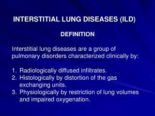
Interstitial lung disease, or diffuse parenchymal lung disease The interstitial lung diseases (ILDs) represent a large number of conditions that involve the parenchyma of the lung—the alveoli, the alveolar epithelium, the capillary endothelium, and the spaces between these structures, as well as the perivascular and lymphatic tissues.
The term "interstitial" is misleading since the pathologic process usually begins with injury to the alveolar epithelial or capillary endothelial cells This heterogeneous group of disorders is classified together because of similar clinical, physiologic, or pathologic manifestations.
classification One useful approach to classification is to separate the ILDs into two groups based on the major underlying histopathology: (1) Those with a predominantly granulomatous reaction in interstitial or vascular areas (2) Those associated with predominant inflammation and fibrosis.
GRANULOMATOUS LUNG DISEASE This process is characterized by an accumulation of T lymphocytes, macrophages, and epithelioid cells organized into discrete structures (granulomas) in the lung parenchyma. The granulomatous lesions can progress to fibrosis. Many patients with granulomatous lung disease remain free of severe impairment of lung function, or, when symptomatic, they improve after treatment.
INFLAMMATION AND FIBROSIS The initial insult is an injury to the epithelial surface causing inflammation in the air spaces and alveolar walls. If the disease becomes chronic, inflammation spreads to adjacent portions of the interstitium and vasculature and eventually causes interstitial fibrosis. .
cont’d The development of irreversible scarring (fibrosis) of alveolar walls, airways, or vasculature is the most feared outcome in all of these conditions because it is often progressive and leads to significant derangement of ventilatory function and gas exchange
Risk groups Age
Gender Smoking history Duration of illness Prior medication use Environmental exposures
CLINICAL PRESENTATION — Patients with ILD commonly come to clinical attention in one of the following ways: • With symptoms of progressive breathlessness with dyspnea or a persistent nonproductive cough. • With an abnormal chest radiograph. • With pulmonary symptoms associated with another disease, such as a connective tissue disease With lung function abnormalities.
LABORATORY STUDIES Serologic studies should be obtained if clinically indicated by : features suggestive of a connective tissue disease (antinuclear antibodies, rheumatoid factor), environmental exposure or systemic vasculitis (antineutrophil cytoplasmic antibodies, anti-glomerular basement membrane antibody).
Chest radiography--- normal in as many as 10 percent of patients with some forms of ILD. Computed tomography--- increasingly important method for the evaluation of diffuse pulmonary parenchymal disease. Serum markers--- including surfactant protein A and B (SP-A, SP-B).
Pulmonary function testing --- Complete lung function testing (spirometry, lung volumes, diffusing capacity) should be obtained. Measurement of lung function is helpful in assessing the severity of lung involvement in patients with ILD.
Three diagnostic techniques are in common use: bronchoalveolarlavage, transbronchial biopsy, and surgical lung biopsy. • Bronchoalveolarlavagemay provide a specific diagnosis in cases of infection, particularly with mycobacteria, or when cytologic examination reveals the presence of malignant cells. • The findings may be suggestive but not diagnostic.
Transbronchial biopsy is easily performed. The risks of pneumothorax and hemorrhage are low. However, the tissue specimens recovered are small, sampling error is common. Transbronchial biopsy can make a definitive diagnosis of sarcoidosis, lymphangitic spread of carcinoma. Transbronchial biopsy cannot establish a specific diagnosis of idiopathic interstitial pneumonia.
Surgical lung biopsy Indications for performing a lung biopsy include the following : To assess disease activity. To exclude neoplastic and infectious processes that occasionally mimic chronic, progressive interstitial disease. To establish a definitive diagnosis and predict prognosis before proceeding with therapies which may have serious side effects