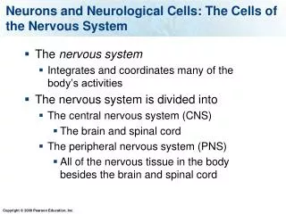
Nervous System Cells
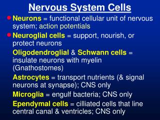
Nervous System Cells. Neurons = functional cellular unit of nervous system; action potentials Neuroglial cells = support, nourish, or protect neurons Oligodendroglial & Schwann cells = insulate neurons with myelin (Gnathostomes)

Nervous System Cells
E N D
Presentation Transcript
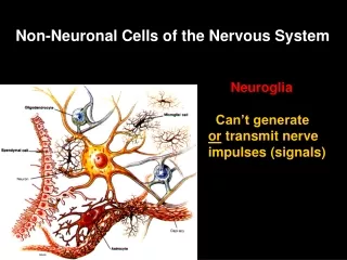
Nervous System Cells • Neurons = functional cellular unit of nervous system; action potentials • Neuroglial cells = support, nourish, or protect neurons Oligodendroglial & Schwanncells = insulate neurons with myelin (Gnathostomes) Astrocytes = transport nutrients (& signal neurons at synapse); CNS only Microglia = engulf bacteria; CNS only Ependymal cells = cilliated cells that line central canal & ventricles; CNS only
Motor Neuron cell body node of Ranvier axon myelin under a Schwann cell axon hillock synaptic terminal dendrite
Neuron Function • Signal propagates down an axon as ion channels open along the axon (changing voltage) – electrical signal • At the end of the axon vessicles containing neurotransmitter release– chemical signal
Subphylum Vertebrata Actinopterygii Chondrichthys coelacanths amphibians lungfishes Mammalia hagfishes lampreys Reptilia myelin (Schwann cells & oligodendrocytes) brain
Nervous System Development • Three ectodermal sources. • Neural tube – becomes C.N.S., optic “nerve,” retina, and pre-ganglionicmotor nerves. CNS derived only from neural tube. • Neural crest – becomes post-ganglionic nerves and peripheral glia. • Neurogenic placodes – become post-ganglionic cranial nerve (V, VII, IX, X) AND olfactory, vestibular, and lateralis sensory receptor cells.
somite notochord inter- mediate meso- derm coelom lateral plate mesoderm Vertebrate Neurula (section) gut
neural crest cells sclerotome dermatome myotome splanchnic mesoderm coelom somatic mesoderm Vertebrate Embryo (section) gut
neural crest cells sclerotome dermatome myotome coelom Vertebrate Embryo (section) gut
neural crest cells vertebra dermatome myotome coelom Vertebrate Embryo (section) gut
will form dorsal root ganglion vertebra dermatome myotome will form other ganglia developing skin coelom Vertebrate Embryo (section) gut
Neurogenic Placodes(post-ganglionic cranial nerves V, VII, IX, & X)
Brain Development • Neurons migrate from the ependymal layer more superficially; 1st migrate least. • The anterior neural tube forms: -Prosencephalon (Forebrain) -Mesencephalon (Midbrain) -Rhombencephalon (Hindbrain) rhombomeres = segmental divisions
Brain Developmental Regions • Prosencephalon = FOREBRAIN Telencephalon cerebrum & olfactory lobes Diencephalon thalamus, hypothalamus • Mesencephalon = MIDBRAIN optic lobes / superior colliculi & tegmentum • Rhombencephalon = HINDBRAIN Metencephalon cerebellum & pons Myelencephalon medulla oblongata
Spinal Nerves Development sensory inter- motor notochord
Nervous System Overview • Central Nervous System (CNS)= brain and spinal cord; myelin from oligodendroglia (astrocytes in synapse) Tract= region of axons; Nucleus or Cortex = region of cell bodies • Peripheral Nervous System (PNS)= nerves and ganglia; myelin from Schwann cells (also can cover synapse) Nerve = bundle of axons Ganglion = bundle of cell bodies
PNS Overview • Cranial Nerves = attached to brain; many have ganglia; numbered with Roman numerals from anterior to posterior • Spinal Nerves = attached to spinal cord; most have ganglia; distinctly segmental dorsal root ventral root
Peripheral Nervous System • Sensory (Afferent) = brings info. to CNS • Motor (Efferent) = takes info. from CNS • Visceral = innervates smooth muscle or organs in the coelom • Somatic = innervates skel. muscle and skin • Pre-ganglionic = cell bodies in the CNS; between CNS & a ganglion • Post-ganglionic Nerves = cell bodies in a ganglion; between a ganglion & another ganglion or a non-neural tissue
Spinal nerves • Spinal Nerves = segmental from spinal cord • Dorsal Root = afferent (sensory) nerve • Dorsal Root Ganglion = afferent (sensory) neuron cell bodies • Ventral Root = efferent (motor) nerve • Sypathetic chain = chain of ganglia ventral to spinal cord
Human Spinal Nerves • Cervical Plexus(C1-C5) also innervates diaphragm (Phrenic nerves) • Brachial Plexus (C5-T1) innervates arm • Intercostal / Thoracic Nerves (T2-T12) • Lumbar Plexus (T12-L5) • Sacral Plexus (L5-Co2) also innervates legs (Sciatic nerves)
dorsal root ganglion dorsal root spinal nerve ventral root sypathetic ganglion Human Thoracic Spinal Nerves
Shingles Herpes/chicken pox virus dormant in spinal or cranial nerve
Cranial Nerves • 10-13 nerves arising from the brain; numbered anterior to posterior. • 0 - nervus terminalis sensory for blood vessels of olfactory epithelium • I - olfactory “nerve” (ethmoid foramina) sensory fibers that innervate the olfactory epithelium • II - optic nerve (optic canal) sensory “nerve;”innervates retina not a true nerve; an extension of brain
Cranial Nerves • III - oculomotor nerve (superior orbital fiss.) motor nerve for 4 of the 6 extrinsic eye muscles • IV - trochlear nerve (superior orbital fissure) motor nerve for 1 of the 6 extrinsic eye muscles • V - trigeminal nerve (3 branches)(foramina ovale and spinosum & superior orbital fissure) sensory for skin of head; motor for 1st arch muscles (mandibular branch) Trigeminal/Gasserion Ganglion • VI - abducens nerve (superior orbital fissure) motor nerve for 1 of the 6 extrinsic eye muscles
Cranial Nerves • VII - facial nerve (3 branches)(stylomastoid foramen) - Facial/Geniculate Ganglion sensory for taste buds and head lateralis system; motor for 2nd arch muscles (hyomandibular branch) • VIII - auditory nerve (Vestibulocochlear) sensory for inner ear/vestibule (internal aud. meatus) • IX - glossopharyngeal nerve (jugular for.) sensory for taste and pharynx; motor for 3rd arch muscles (1st branchial arch branch) – Superior & Inferior Glossopharyngeal Ganglia • IX - vagus nerve (jugular foramen) sensory and motor for mouth, pharynx, outer ear, and most viscera – Superior & Inferior Vagus Ganglia
Posterior-Most Cranial Nerves • XI - spinal accessory nerve (amniotes only) (foramen magnum) sensory & motor for branchiomeric muscles (e.g., trapezius, sternomastoid) = branch of vagus in non-amniotes • XII - hypoglossal nerve (amniotes only) (hypoglossal canal) motor & sensory for tongue muscles = occipital nerves in non-amniotes
somitomeres 1-7 somites 1-4 mandibular arch hyoid arch 1st branchial arch Shark-like Vertebrate
terminal 0 mandibular arch hyoid arch 1st branchial arch Special Sensory Cranial Nerves optic II auditory VIII olfactory I
X XII IV V VI VII IX III mandibular arch hyoid arch 1st branchial arch Segmental Cranial Nerves XI
X XII IV V VI VII IX III mandibular arch hyoid arch 1st branchial arch Segmental Cranial Nerves XI II VIII I 0
X XII VII IV VI V IX III XI II 0 I VIII Suprabranchial Placodes • Form ganglia for the trigeminal, facial, glossopharyngeal, & vagus cranial nerves.
CNS • Spinal Cord = CNS dorsal to notochord and posterior to head • Central canal = canal within spinal cord • Brain = enlarged anterior CNS • Ventricles = cavities in brain • Cerebrospinal fluid (CSF) = fills ventricles & central canal • Choroid plexus = tuft of capillaries that secrete CSF
Meninges • Meninges = layers of connective tissue surrounding CNS Non-tetrapods = 1 (primitive meninx) Non-mammal tetrapods = 2 (dura matter + secondary meninx) Mammals = 3 • Dura matter = outermost • Arachnoid = middle • Pia matter = innermost
CNS • Gray matter = many cell bodies; >integration • White matter = few cell bodies; many axons with myelin(in Gnathostomes); >transport • Gray matter ancestrally deep; white matter superficial, EXCEPTsuperficial in forebrain in amniotes, teleosts, hagfishes, and a few chondrichthyans
Spinal Cord cauda equina
Brain Regions • Prosencephalon = FOREBRAIN Telencephalon cerebrum & olfactory lobes Diencephalon thalamus, hypothalamus, epiphysis, hypophysis, optic nerves • Mesencephalon = MIDBRAIN; tectum superior colliculi & inferior colliculus tegmentum • Rhombencephalon = HINDBRAIN Metencephalon cerebellum & pons Myelencephalon medulla oblongata
Teleost Brain Regions Pros- Mes- Rhomb-
Bird Brain Regions Pros- Mes- Rhomb-
Telencephalon • Olfactory lobes • Cerebrum, large in mammals & birds, lateral ventricles. • In mammals, dorsal pallium forms the 6 layered neocortex. • gyri = folds of neocortex • sulci = grooves of neocortex • Actinopterygiians = gray matter everts; single medial ventricle • All other vertebrates = gray matter inverts; 2 lateral ventricles
Diencephalon • Thalamus, Hypothalamus, & Median Ventricle (3rd). • Optic nerves cross and enter brain at optic chiasma (neurons pass through the diencephalon, synapse in the mesencephalon). • Dorsal epiphysis (pineal & parietal organ) & Ventral hypophysis (pituitary).