Download
1 / 87
880 likes | 1.11k Vues
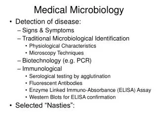
REVIEW OF MEDICAL MICROBIOLOGY. Infections of Respiratory tract Cardiovascular system Gastrointestinal tract Skin and soft tissue Central nervous system Genitourinary tract. THE RESPIRATORY TRACT. Upper Respiratory Tract Pharyngitis (mostly 2 years through adolescence)

E N D
REVIEW OF MEDICAL MICROBIOLOGY Infections of Respiratory tract Cardiovascular system Gastrointestinal tract Skin and soft tissue Central nervous system Genitourinary tract
THE RESPIRATORY TRACT Upper Respiratory Tract Pharyngitis (mostly 2 years through adolescence) Adenoviruses Group A Streptococci (S. pyogenes) Potential for rheumatic fever Chlamydophila pneumoniae Neisseria gonorrhoeae Corynebacterium diphtheriae Mycoplasma pneumoniae
THE RESPIRATORY TRACT Otitis media (infants and young children) Streptococcus pneumoniae Haemophilus influenzae Staphylococcus aureus Group A streptococcus Moraxella catarrhalis Formerly “Branhamella” Gram-negative cocci Opportunistic pathogen
THE RESPIRATORY TRACT Otitis externa Staphylococcus aureus Pseudomonas aeruginosa Group A Streptococcus Malignant otitis externa • In diabetics, elderly & immunocompromised • Can lead to osteomyelitis and meningitis
THE RESPIRATORY TRACT Sinusitis Streptococcus pneumoniae Haemophilus influenzae Staphylococcus aureus Chlamydophila pneumoniae Moraxella catarrhalis Group A Streptococcus Pseudomonas aeruginosa Viruses Oral anaerobic bacteria
THE RESPIRATORY TRACT Conjunctivitis Streptococcus pneumoniae Group B Streptococcus Viridans Streptococcus Staphylococcus aureus Haemophilus influenzae Moraxella catarrhalis
THE RESPIRATORY TRACT Conjunctivitis (contd) Pseudomonas aeruginosa Corynebacterium species Francisella tularensis Adenoviruses Chlamydia trachomatis
THE RESPIRATORY TRACT Rhinocerebral mucormycosis • Life-threatening • Most common in diabetics • The fungi Mucor and Rhizopus invade blood vessels, resulting in necrosis of bone and thrombosis of the cavernous sinus and internal carotid artery
THE RESPIRATORY TRACT Bacterial epiglottitis Life-threatening Haemophilus influenzae type b Streptococcus pneumoniae Staphylococcus aureus
THE RESPIRATORY TRACT Diphtheria Corynebacterium diphtheriae Whooping cough Bordetella pertussis
THE RESPIRATORY TRACT “Common colds” Rhinoviruses Adenoviruses Influenza C Coronaviruses Coxsackie viruses
THE RESPIRATORY TRACT “Croup” Respiratory syncytial virus Influenza virus Parainfluenza virus
THE RESPIRATORY TRACT Lower Respiratory Tract Community acquired infections Streptococcus pneumoniae (elderly) Klebsiella pneumoniae (alcoholics) Mycoplasma pneumoniae (school-age children) Mycobacterium tuberculosis RSV (infants and young children) Influenza virus
THE RESPIRATORY TRACT Lower Respiratory Tract Community acquired infections Bronchitis or pneumonia secondary to viral pneumonia Streptococcus pneumoniae Haemophilus influenzae Staphylococcus aureus Moraxella cararrhalis
THE RESPIRATORY TRACT Lower Respiratory Tract Nosocomial infections Mycobacterium tuberculosis RSV in pediatric patients Methicillin-resistant S. aureus (pneumonia) Pseudomonas aeruginosa Legionella spp.
THE RESPIRATORY TRACT Lower Respiratory Tract Patients with underlying lung infections Chronic obstructive pulmonary disease P. aeruginosa S. pneumoniae H. influenzae Moraxella cararrhalis Allergic bronchopulmonary aspergillosis
THE RESPIRATORY TRACT Lower Respiratory Tract Patients with underlying lung infections Cystic fibrosis S. aureus P. aeruginosa Allergic bronchopulmonary aspergillosis
THE RESPIRATORY TRACT Lower Respiratory Tract Patients with underlying lung infections Cavitary lung disease (due to prior MTB infection) Aspergillus spp (Aspergilloma or fungus ball)
THE RESPIRATORY TRACT Lower Respiratory Tract Immunocompromised individuals At risk for all recognized respiratory tract pathogens AIDS patients Pneumocystis carinii S. pneumoniae MDR M. tuberculosis
THE RESPIRATORY TRACT Lower Respiratory Tract Immunocompromised individuals Neutropenic patients Invasive aspergillosis Mucormycosis
THE RESPIRATORY TRACT Lower Respiratory Tract Immunocompromised individuals Transplant patients Invasive fungi CMV HSV Legionella spp. Pneumocystis carinii
A 40-year-old male with multisystem failure secondary to bilateral pneumonia was transferred to our hospital via helicopter. He had presented to his local physician 3 days previously complaining of fever, malaise, and vague respiratory symptoms. He was given amantadine for suspected influenza. His condition became progressively worse, with shortness of breath and a fever to 40.5˚C. From: “Cases in Medical Microbiology and Infectious Disease”
He was admitted to an outside hospital 24 h prior to transfer. A laboratory examination revealed abnormal liver and kidney function. Therapy with Timentin (ticarcillin-clavulanic acid) and trimethoprim-sulfamethoxazole was begun. He underwent pronchoscopic examination which revealed mildly inflamed airways containing thin, watery secretions.
A Gram-stain of bronchial washings and culture results are shown in the figure. Based on these findings, he was begun on appropriate antimicrobial therapy.
Which organisms are common causes of community-acquired bacterial pneumonia?
Streptococcus pneumoniae Haemophilus influenzae Mycoplasma pneumoniae Staphylococcus aureus (frequently following an influenza infection) Klebsiella pneumoniae (elderly & alcoholics) Legionella pneumophila Chlamydophila pneumoniae
On the basis of the Gram-stain of bronchial washings, and the patient’s presentation, what is the most likely cause of this patient’s catastrophic infection? Why must the laboratory be notified if this organism is considered in the differential diagnosis?
The patient has Legionella pneumophila. Renal and hepatic dysfunction and thin watery secretions are characteristic of this infection. Patients with bacterial pneumonia due to most other bacterial agents have thick, purulent secretions. The laboratory needs to be informed because the organism requires a specific growth medium, buffered charcoal yeast extract (BCYE) agar.
What techniques other than culture can be used to detect this organism within 24 h?
What is the appropriate antimicrobial agent for the treatment of this infection? Which other Gram-negative respiratory pathogen is treated with this antibiotic?
Erythromycin Can penetrate into white blood cells Legionella multiplies in macrophages Bordetella pertussis
THE CARDIOVASCULAR SYSTEM Septicemia: Predisposing factors and agents Abdominal sepsis Enterobacteria Bacteroides fragilis Enterococcus faecalis Enterococcus faecium Infected wounds Staphylococcus aureus Streptococcus pyogenes Enterobacteria
THE CARDIOVASCULAR SYSTEM Septicemia: Predisposing factors and agents Osteomyelitis Staphylococcus aureus Pneumonia Streptococcus pyogenes Food poisoning Salmonella spp. Campylobacter spp.
THE CARDIOVASCULAR SYSTEM Septicemia: Predisposing factors and agents Intravascular devices Staphylococcus aureus Staphylococcus epidermidis Enterobacteria Meningitis Streptococcus pneumoniae Neisseria meningitidis Haemophilus influenzae
THE CARDIOVASCULAR SYSTEM Septicemia: Predisposing factors and agents Immunocompromised patients Staphylococcus aureus Enterobacteria
THE CARDIOVASCULAR SYSTEM Infective endocarditis > 80% of cases caused by streptococci or staphylococci Total streptococci 60% Viridans group 35% anginosus group mitis group mutans group salivarius group
THE CARDIOVASCULAR SYSTEM Infective endocarditis Total streptococci 60% Total staphylococci 25% S. aureus 20% S. epidermidis 5%
THE CARDIOVASCULAR SYSTEM Myocarditis Corynebacterium diphtheriae Clostridium perfringens Group A Streptococcus Borrelia burgdorferi Neisseria meningitidis Staphylocccus aureus
The patient was a 4-month-old female who was admitted to the hospital in March with sever respiratory distress. Five days prior to admission she had developed a cough and rhinitis. Two days later she began wheezing and was noted to have a fever. She was brought to the emergency room when she became lethargic. From: “Cases in Medical Microbiology and Infectious Disease”
One sibling was reported to be coughing, and her father had a “cold”. On examination she had a fever of 38.9˚C tachycardia with a pulse of 220/min tachypnea with respirations of 80/min Her throat was clear.
A chest X-ray revealed interstitial infiltrates. She was put in respiratory isolation in the pediatric intensive care unit, and was subsequently intubated. Blood and nasopharyngeal cultures were sent to the bacteriology and virology laboratories. A rapid diagnostic test was positive and specific antiviral therapy was begun.
She was also given a bronchodilator (aminophylline) to treat the bronchospasm which was resulting in her wheezing. She was extubated 5 days later and discharged home on day 8. 1. What are the possible causes for this patient’s pneumonia?
Parainfluenza virus Influenza A and B Respiratory syncytial virus Mycoplasma pneumoniae Bordetella pertussis
2. What other techniques could one use to identify this microorganism?
Direct Fluorescence Antibody “Shell Vial Assay” Fibroblasts grown on coverslips in a shell vial Clinical specimens a centrifuged onto the cell monolayer Incubation for 1-2 days The monolayer is stained with a fluorescent monoclonal antibody specific for an RSV antigen
RSV is spread by large droplets and on fomites Can be spread via contaminated hands Occurs primarily in winter months